Site sections
Editor's Choice:
- The best recipes for tomato paste for the winter at home
- How to cook pea soup with smoked ribs
- Luxurious custard dough for yeast pies
- Pumpkin porridge with rice in a slow cooker
- Amber transparent jam from apple slices for the winter
- Salad "Krasnaya Polyana" Salad "Krasnaya Polyana"
- Nigiri - sushi recipe at home Nigiri sushi at home
- Harvesting mushrooms for the winter - recipes
- Sweet omelet with semolina
- Egg and milk casserole
Advertising
| What is the excretory system of reptiles. quick lizard |
|
The heart of lizards is three-chambered, has two atria and one ventricle, divided into three parts: the venous cavity, the arterial cavity and the lung cavity. The venous cavity receives oxygen-poor blood from the right atrium, and the arterial cavity receives oxygen-rich blood from the left atrium. Blood leaves the heart through the pulmonary artery, originating in the lung cavity and two aortic arches, extending from the venous cavity. All three lizard heart cavities communicate, but the muscle flap and biphasic ventricular contraction minimize blood mixing. Oxygen-poor blood flows from the venous cavity into the pulmonary cavity, the atrioventricular valve prevents it from mixing with oxygen-rich blood from the arterial cavity. The contraction of the ventricle then pushes this blood out of the lung cavity into the pulmonary artery. The atrioventricular valve then closes, allowing oxygen-rich blood from the arterial cavity to enter the venous cavity and leave the heart through the aortic arches. Thus, a three-chambered heart is functionally similar to a four-chambered one. The paired left and right aortic arches merge behind the heart into the dorsal aorta. Reptiles have a portal system of the kidneys - the venous vessels of the tail and partly of the hind limbs lead directly to the kidneys. Thus, if tubular filtration drugs are injected into the posterior half of the body, their serum concentrations may be lower than expected due to premature urinary excretion. In the case of the introduction of nephrotoxic drugs, side effects may increase. However, there have been few studies of this effect and their results indicate rather an insignificant role of the renal portal system in pharmacokinetics. Moreover, there are shunts in the system that carry blood from the renal portal system to the posterior vena cava, bypassing the renal tissue. Lizards have a large abdominal vein that lies along the inner surface of the middle of the abdominal wall on a ligament a few millimeters from the white line. When performing abdominal operations, this vein is tried to be avoided. However, if damaged, it can be ligated without complications. Digestive system of lizards Genitourinary system of lizards Nervous system of lizards Sense organs of lizards Respiratory system of lizards Musculoskeletal system of lizards Endocrine system of lizards CLASS REPTILIA - REPTILIA TOPIC 12. OPENING THE LIZARD SYSTEMATIC POSITION OF THE OBJECT Subtype Vertebrates, Vertebrata MATERIAL AND EQUIPMENT For one or two students you need: EXERCISE Get to know the features of the external appearance of the lizard. Pay attention to the parts of the body, the structure of the integument, external structure eye, nostril openings, ear openings, etc. Perform an opening. Familiarize yourself with the general arrangement of internal organs; sequentially consider the structure of individual organ systems, starting with the circulatory system. Make the following drawings: Additional task Examine, without sketching, under a microscope, a section of the skin of a lizard. APPEARANCE The body of the lizard is clearly divided into the head, neck, trunk, tail and paired limbs - front and rear (Fig. 71). Rice. 71. Appearance (A) and the area of the cloaca from below (B) of the Caucasian agama, male: The surface layers of the epidermis of the skin of the lizard (as well as all other reptiles) become keratinized: the cells gradually die off, being filled with a horny substance - keratohyalin. The thickening of the stratum corneum occurs in small areas - scales, between which the stratum corneum is very thin (Fig. 72), so the flexibility of the skin (and the whole body) is preserved. The shape of the scales on different parts of the body of the same animal can vary significantly. In different species, the shape, arrangement and number of scales are usually more or less different, therefore these features are widely used in the taxonomy of reptiles.
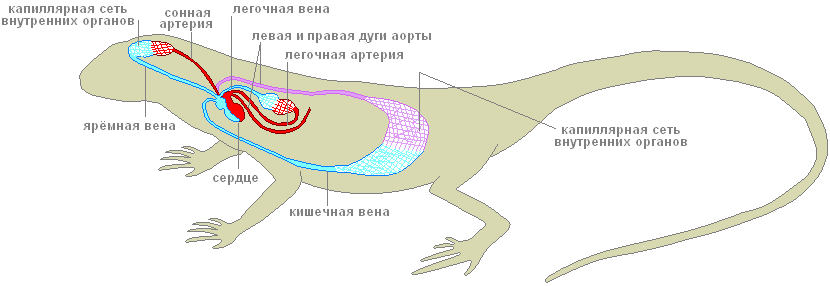
Rice. 72. Scheme of a cross section of the skin of a lizard of the genus Lacerta: The head of the agama is covered with small, irregular shape scales; some other lizards (for example, the genera Lacerta, Eremias) have rather large horny scutes on their heads, arranged in a strictly defined order. On the upper surface of the head, paired external nostrils are visible (Fig. 71, 1), opening into the oral cavity with the so-called internal nostrils, or choanae (check by introducing a needle or bristle!). The eyes (Fig. 71, 2) are covered with movable eyelids; there is a nictitating membrane in the back corner of the eye. Behind the eyes are ear openings (Fig. 71, 3), at some depth tightened by the tympanic membrane. The elongated body of the agama is also covered with horny scales (Fig. 71, 5) - small, irregularly shaped scales on the dorsal side and rows of larger scutes on the belly. At the posterior end of the body, on the border with the caudal region, between the abdominal shields, there is a slit-like opening of the cloaca (Fig. 71, 6). The tail scales of the Caucasian agama form double rings; in other lizards, the location of the tail scales is different. The five-fingered limbs of lizards, like other reptiles, end in horn formations - claws (Fig. 71, 4). The skin of lizards, like all reptiles, is dry, which is associated with the absence of mucous glands. Skin glands are present in small numbers and are located only in a few areas defined for this species. They secrete a thick fat-like secret and have special functions, most likely associated with leaving an odorous trail that facilitates the formation of pairs during reproduction. In the agama, a group of such glands is clearly visible in the back of the abdomen; their secret in the form of a "wax" coating covers the scales in this area. This accumulation of glands is especially well expressed in males. OPENING 1. Place the lizard on its back in the wax bath and pin the limbs to the bath. GENERAL TOPOGRAPHY OF THE INTERNAL ORGANS Circulatory system. The heart (cor) is located on the ventral side of the front of the chest cavity. Like amphibians, the heart of lizards is three-chambered: it consists of two atria - right and left (atrium dexter et atrium sinister; Fig. 73, 1, 2) and one ventricle (ventriculus; Fig. 73, 3).
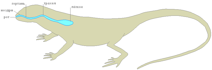
Rice. 73. Scheme of the circulatory system of the Caucasian agama The ventricle of the heart is divided by an incomplete, so-called horizontal septum into two cavities: a smaller ventral (more precisely, ventrolateral), located down and to the right of the septum, and a large dorsal (dorsolateral) - up and to the left of the septum. The left atrium opens into the left side of the dorsal cavity of the stomach, and the right atrium opens into right side the same cavity, in the region of the free edge of the septum. The dorsal cavity is divided into separate chambers by numerous muscular ridges. One of them, the most developed, is the so-called vertical septum, which divides the dorsal cavity of the ventricle into two halves - left and right. Due to this structure, complete mixing of arterial and venous blood does not occur in the ventricle of the heart of reptiles. During atrial contraction, arterial blood expelled from the left atrium collects mainly in the left side of the dorsal cavity of the ventricle; venous blood from the right atrium enters the right half of the dorsal part of the ventricle and, flowing around the edge of the horizontal septum, is collected in the ventral part of the ventricle. Only in the right half of the dorsal part of the ventricle does the arterial and venous blood mix. The arterial cone characteristic of amphibians in reptiles is reduced, and the main arterial trunks of the systemic and pulmonary circulations depart from the ventricle on their own. At the same time, unlike amphibians, in which three pairs of arterial trunks depart from the arterial cone, in reptiles only three unpaired vessels begin in the heart: the pulmonary artery and two (right and left) aortic arches. The pulmonary artery (arteria pulmonalis; Fig. 73, 4) starts from the ventral (venous) part of the ventricle and soon divides into two branches that carry blood to the right and left lungs. Venous blood moves through the pulmonary arteries. Oxygenated arterial blood through the pulmonary veins (vena pulmonalis; Fig. 73, 5) returns to the heart. The right and left pulmonary veins merge into one unpaired vessel that flows into the left atrium. The entire system of the considered vessels constitutes a small (pulmonary) circle of blood circulation. The vessels of the systemic circulation also begin in the ventricle of the heart. The right aortic arch (arcus aortae dexter; Fig. 73, 6) departs from its left dorsal (arterial) part, and to the right of it, in the region of the free edge of the horizontal septum, the left aortic arch (arcus aortae sinister; Fig. 73, 7) . According to the place of origin of these vessels in the ventricle, predominantly arterial blood enters the right aortic arch, while mixed blood enters the left arch (arterial with an admixture of venous). Both aortic arches go around the heart and on the dorsal side behind it are combined into an unpaired dorsal aorta (aorta dorsalis; Fig. 73, 8), which sends numerous vessels to various organs of the body. In the region of the hind limbs, the dorsal aorta branches into two large iliac arteries (arteria iliaca; Fig. 73, 9), which carry blood to the limbs, and the tail artery (arteria caudalis; Fig. 73, 10). The carotid arteries (arteria carotis; Fig. 73, 11) depart from the right aortic arch with a short, immediately bifurcating common trunk. Both carotid arteries, initially running parallel to the ascending branches of the aortic arches, carry blood to the head above the place where the aortic arches turn upwards (downward from the observer) and backwards, each carotid artery sends out the carotid duct (ductus caroticus; Fig. 73, 12), which flows respectively into the right or left aortic arch. All of these vessels are quite clearly visible on a freshly killed lizard. If you carefully dissect the right aortic arch, then approximately in the middle between the place of its turn and the place of confluence of the aortic arches, at the level of the posterior end of the heart, you can see the subclavian arteries (arteria subclavia; Fig. 73, 13) extending from it, going to the forelimbs. Thus, in reptiles, unlike amphibians, the carotid and subclavian arteries depart asymmetrically - only from the right aortic arch. Thanks to this, blood, the richest in oxygen, enters the head and forelimbs. Venous blood from the head is collected in large paired jugular veins (vena jugularis; Fig. 73, 14), which, merging with less visible subclavian veins (vena subclavia; Fig. 73, 15) coming from the forelimbs, form paired anterior vena cava ( vena cava anterior dextra et vena cava anterior sinistra; Fig. 73, 16). The anterior vena cava flows into the venous sinus (sinus venosus; Fig. 73, 17), which communicates with the right atrium. In lizards, the venous sinus, like in most reptiles, is weakly expressed. From the back of the body, venous blood enters the heart in two ways. The veins that carry blood from the hind limbs form short paired portal veins of the kidneys (vena porta renalis; Fig. 73, 18), with each of which the branches of the divided unpaired tail vein (vena caudalis; Fig. 73, 19) merge. These vessels can usually be seen only on injected preparations. Through the portal veins of the kidneys, blood enters the capillary system - the portal system of the kidneys. Most of the blood from the posterior part of the body goes through rather large paired pelvic veins (vena pelvica; fig. 73, 20; sometimes they are called iliac veins - v. iliaca), which, merging, form an unpaired abdominal vein (vena abdominalis; fig. 73, 21), which carries venous blood to the liver. Venous blood from the intestines goes through several veins that merge into the unpaired portal vein of the liver (vena porta hepatis; Fig. 73, 22). In the liver or before entering it, the portal vein of the liver merges with the abdominal vein, and this common vessel immediately breaks up into a system of hepatic capillaries. Consequently, as in amphibians, the portal system of the liver is formed by two veins: the abdominal and portal liver. From the portal system of the kidneys, blood is collected in the paired renal veins (vena renalis; Fig. 73, 23), which merge into a large unpaired posterior vena cava (vena cava posterior; Fig. 73, 24). The posterior vena cava pierces the liver (without sending vessels to it) and flows into the venous sinus. From the portal system of the liver, blood is collected through the capillary system into a short hepatic vein (vena hepatica; Fig. 73, 25), which flows into the posterior vena cava in the region of the anterior edge of the liver. Respiratory system. The respiratory tract of the lizard begins with external nasal openings - the nostrils. Further, the air through the nasal passage and internal nostrils - choanae, enters the oral cavity. In the depths of the oral cavity, somewhat in front of the esophagus, there is a larynx (larynx), consisting of three cartilages. It is equipped with special muscles and is connected with the sublingual apparatus. From the oral cavity, the inhaled air through the larynx enters the trachea (trachea; Fig. 74, 4) - a rather long tube, in the walls of which there are ring-shaped cartilages that prevent it from collapsing. The trachea runs along the neck and in the chest cavity, approximately at the level of the heart, is divided into two short bronchi (bronchus), which enter the lungs. Lungs (pulmones; Fig. 74, 5) are thin-walled hollow bags. Compared to the lungs of amphibians, lizards have a more complex internal structure: their inner walls, in which capillaries branch, have a spongy structure, which significantly increases the total respiratory surface of the lungs. The lungs are the only respiratory organ in reptiles. The skin of these animals is dry, covered with horny scales and keratinized epithelium, and does not participate in respiration. The act of breathing in lizards occurs by expanding and contracting the chest under the action of special muscles.
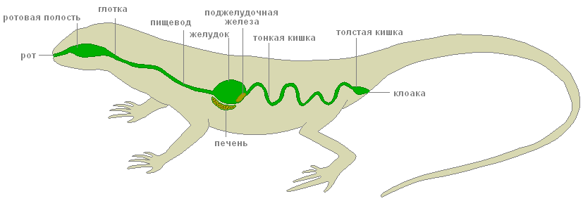
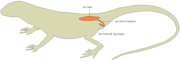
Rice. 74. General arrangement of the internal organs of the female Caucasian agama: Digestive system. In the oral cavity is a flat, anteriorly tapering tongue; it aids in the capture and swallowing of prey. Many lizards and snakes have a thin and long tongue that forks at the end. It is very mobile, can protrude quite far from the mouth and also performs the function of an organ of touch: lizards and snakes feel objects in front of them. In addition, when the tongue is retracted into the mouth, its tips fall into special depressions equipped with sensory nerve endings - the Jacobson organ, which perceives chemical irritations from particles adhering to the tongue. At the posterior end of the oral cavity behind the laryngeal fissure is the opening of the esophagus. The esophagus (oesophagus; Fig. 74, 6) in the form of a muscular tensile tube stretches along the neck above the trachea and flows into the stomach in the anterior part of the abdominal cavity (gaster; Fig. 74, 7). From the posterior end of the stomach forward parallel to it is the duodenum (duodenum; Fig. 74, 8), passing into the small intestine (ileum; Fig. 74, 9). The border of the duodenum and small intestine is the first bend of the intestine (the place where the intestine turns back). The small intestine makes several bends and passes into the large intestine (colon; Fig. 74, 10). On the border of the small and large intestines there is a small blind outgrowth - the rudiment of the caecum (coecum; Fig. 74, 11). The posterior part of the large intestine is the rectum (rectum; Fig. 74, 12). In lizards, the colon and rectum are separated by a poorly visible narrowing. The rectum opens into the cloaca (cloaca; Fig. 74, 13) and out through the cloacal fissure. Between the stomach and duodenum is an elongated compact pancreas (pancreas; Fig. 74, 14). Near the stomach, towards the end of it, there is a small elongated reddish (on fresh material) spleen (lien; Fig. 74, 15). The entire anterior part of the abdominal cavity (behind the heart) is occupied by a large liver with several lobes (hepar; Fig. 74, 16). On her inside the gallbladder (vesica fellea; fig. 74, 17) is located. The bile duct departing from it (ductus choledochus; Fig. 74, 18) runs along the pancreas and flows into the beginning of the duodenum. The bile duct becomes more visible if you lightly press the tweezers on the gallbladder and thus push some of the bile into the duct. Urogenital system. In contrast to the previously studied classes, adult reptiles function not in the trunk (mesonephric), but in the pelvic (metanephric) kidneys (ren; fig. 75, 1; fig. 76, 1). They are located in the posterior part of the abdominal cavity and are covered by the bones of the pelvis. Along each kidney is the ureter (ureter), which opens into the cloaca. The ureters of lizards, like those of other reptiles, are formed simultaneously with the development of the metanephric kidney as thin-walled protrusions of the posterior part of the wolfian canals. The urinary bladder (vesica urinaria; Fig. 75, 2; Fig. 76, 2) departs from the abdominal wall of the cloaca in the form of a thin-walled blind outgrowth.
Rice. 75. Urogenital system of a male Caucasian dragon: The sex glands of males - paired testes (testis; Fig. 75, 3) - are suspended on the mesentery in the posterior dorsal part of the abdominal cavity. The testes, with the help of the seminiferous tubules, are closely connected with the appendages of the testes (epididymis; fig. 75, 4), from which the vas deferens (vas deferens; fig. 75, 5) go. Just before flowing into the cloaca, the vas deferens merge with the ureters and open in the cloaca with common openings (Fig. 75, 6). The appendages of the testis are the remains of the anterior part of the trunk (mesonephric) kidney, and the vas deferens are homologous to the excretory duct of this kidney - the wolfian canal. Müllerian canals do not develop in males. In the side walls of the cloaca in males there are two hollow outgrowths that can turn outward through the opening of the cloaca. They play the role of copulatory organs. Rice. 76. The genitourinary system of a female Caucasian agama: The sex glands of females are paired ovaries (ovarium; Fig. 76, 4), suspended in the abdominal cavity on the mesentery and have no direct connection with the excretory ducts. Mature eggs fall into the body cavity and then are captured by the funnel of the oviduct (Fig. 76, 6), which opens in front of the body cavity. The oviducts (oviductus; Fig. 76; 5), homologous to the Müllerian canals, open into the cloaca with independent (separate from the ureters) openings (Fig. 76, 7). The lower sections of the oviducts in lizards are often enlarged and then they are called the "womb". Wolf channels in females are reduced. General characteristics of the class Watch the lecture. Features of the organization of reptiles The body shape of reptiles is very different, which is associated with a variety of modes of movement. All parts of the body are expressed : head, body, tail. Turtles have a body more or less flattened in the dorsal-abdominal direction and enclosed in a shell. covers reptiles differ significantly from the integument of amphibians. The upper layers of the multilayered epidermis are keratinized; cells are filled with keratin protein, the grains of which displace the protoplasm and nucleus. The skin of reptiles has lost the ability to exchange gases, evaporate water and excrete metabolic products. The skin of reptiles is practically devoid of skin glands, so numerous in amphibians. The change of the horny cover is provided by full or partial molting, which in some species may occur several times a year. Skeleton. The axial skeleton of reptiles is represented by the spine, in which, unlike amphibians, 5 departments: cervical, thoracic, lumbar (appears for the first time), sacral and caudal. In the cervical region, the number of vertebrae is 7-10. A feature of this section of the axial skeleton is not only a greater number of vertebrae compared to amphibians, but also differentiation first two vertebrae: first cervical vertebra - atlas or atlas ( atlas) - has the form of a bone ring, divided by a dense ligament into the upper and lower halves. The upper opening serves to connect the brain with the spinal cord, the odontoid process of the second cervical vertebra, the epistrophy, enters the lower one ( epistropheus). To the vertebrae thoracic(16-25 sterno-lumbar vertebrae) ribs are attached, connecting to the sternum with their abdominal ends, forming closed chest characteristic of most reptiles. The belt of the forelimbs is also attached to the sternum. Vertebrae lumbar bear ribs that do not reach the sternum. sacral department represented by two vertebrae, to the transverse processes of which the ilium bones of the pelvic girdle are attached. tail section consists of 15-40 vertebrae, performs various functions : helps to maintain balance when moving, serves as a mover (for sea snakes, crocodiles, water lizards). In lizards capable of autotomy, each tail vertebra can break in the middle where the cartilaginous layer is located, dividing the vertebral body into two parts. Paired limbs and their belts. Shoulder girdle reptiles consists mainly of the same elements as those of amphibians, but most of its elements are ossified. Pelvic girdle consists of two nameless bones, each of which is represented by three bones : iliac, sciatic and pubic, forming the acetabulum, which forms a joint with the femoral head. Paired limbs basically correspond to the plan of the structure of the limbs of terrestrial vertebrates. Scull reptiles is distinguished, first of all, by complete ossification and development a large number cover bones. musculature. The metameric structure was preserved only by the muscles connecting the adjacent vertebrae and the muscles of the abdominal wall. Digestive organs and nutrition. Modern reptiles - predominantly carnivores. The capture and retention of prey is carried out by jaws with numerous sharp teeth located on them. The teeth of reptiles are not differentiated; some species of snakes develop large poisonous teeth. Reptile prey, as a rule, is swallowed whole, only crocodiles and turtles are able to tear off individual pieces from large prey. The special device of the jaw apparatus of snakes allows them to swallow prey that exceeds the normal width of snakes. In the oral cavity of reptiles are located salivary glands(there are enzymes, but not enough). In poisonous snakes and lizards, some of the salivary glands have become poisonous. At the bottom of the oral cavity is a movable muscular tongue that can extend far. The esophagus is well defined. The stomach is separated from the esophagus, has muscular walls, passes into the intestines. The intestine opens into the cloaca. The pancreas lies in the first loop of the intestine. The large liver of reptiles has a gallbladder, the duct of which flows into the intestine next to the pancreatic duct. Feature of functioning digestive system reptiles indicate that this is a heat-loving group of animals. Digestion of large prey, for example, in snakes, proceeds normally only at a sufficiently high temperature (+ 20-23 C) ; slowing down digestion at low temperatures causes food poisoning or entails belching of prey. The ability of reptiles, especially snakes and turtles, to long-term starvation (up to 2 years in captivity) is amazing. Respiratory organs and gas exchange. The skin of reptiles does not take part in respiration, and paired lungs serve as the main respiratory organs of reptiles. The general form of light reptiles, like amphibians, is bag-shaped, however internal structure much more difficult. In turtles and crocodiles, the lungs have a spongy structure, reminiscent of the structure of the lungs of birds and mammals. Ventilation of the lungs is provided by the work of the chest with the help of the intercostal and abdominal muscles. The circulatory system and circulation. The heart of reptiles, like that of amphibians, three-chamber. The atria are separated by a complete septum; each opens into the ventricle with an independent opening, equipped with a valve of semilunar folds. The ventricle has an incomplete septum extending from its ventral side and dividing it into two parts : at the time of systole, the septum reaches the dorsal wall of the ventricle, a short time separating it, which is important for separating blood streams with different oxygen content. In crocodiles, this septum is complete, but with a hole in the center. The venous sinus is fused with the right atrium. The arterial cone is reduced. excretory organs reptiles represented pelvic- metanephric - by the kidneys. The end products of nitrogen metabolism are several substances - ammonia, uric acid, urea and others, but, as a rule, any one predominates. The metanephric (pelvic) kidney differs not only in its position (located in the pelvic region), but also in the complication of the structure of the renal (nephron) tubules. As a result, 90-95% of the primary filtrate returns to the bloodstream. The end urine enriched with waste products flows from the kidneys through the ureters to the cloaca and bladder, where water reabsorption is completed, after which the concentrated urine is excreted from the body. In reptiles, in the process of evolution, the need arose to conserve water. Reproductive organs represented by paired sex glands. The testicles have appendages, which are the remnant of the mesonephric kidney of the embryos. The right and left vas deferens (they are the ducts of the mesonephric kidney, i.e. the Wolfian channels), coming from the testes, open into the corresponding ureters at their confluence with the cloaca. Fertilization is only internal. Paired ovaries look like oval granular bodies. The oviducts are the Muller canals. Fertilization occurs in the upper part of the oviduct. In the middle section of the oviduct, there are glands that form around the egg the protein shell of the egg, which is poorly developed in snakes and lizards and powerful in crocodiles and turtles. In the walls of the lower part of the oviduct (uterus) there are glands that form a parchment-like or lime-soaked shell of the egg. Most reptiles bury the eggs they lay in the ground in well-warmed places; some species lay their eggs in heaps of plant humus or under rotting stumps. Lisher masonry (lizards, etc.), snakes guard. The fecundity of reptiles is much lower than that of amphibians. A few modern representatives of the neg. Scaly exists ovoviviparous or less commonly viviparity. Nervous system and sense organs. There are 5 parts of the brain. More developed than amphibians. There are 11 pairs of head nerves. The organ of vision of reptiles is adapted to work in the air. The eye is protected by the outer eyelids and the nictitating membrane. In snakes and some lizards, the eyelids fuse together to form a transparent shell. Most reptiles have developed color vision. The organ of hearing, as in amphibians, is represented by the inner and middle ear with the tympanic membrane, containing one auditory bone - the stirrup. Most reptiles nemo; loud roaring sounds are made by crocodiles, the sounds of snakes are represented by hissing, wheezing, and the sound of tail rattles. All of these sounds serve basically as a menacing warning. Chemoreception also plays an important role in the orientation and communication of reptiles. The olfactory organs open outward through paired nostrils. In the roof of the oral cavity there is a recess, the so-called Jacobson organ, which perceives the smell of food or substances in the mouth, which the animal collects from the ground with a movable tongue and directs into the oral cavity. Some reptiles (snakes, pythons, African vipers) have special organs of thermal sense, represented by thermoreceptors and even thermolocators (pit vipers). The sense of touch in reptiles is also pronounced. population organization reptiles are harder than amphibians. Most reptiles lead a solitary lifestyle during the active season. During hibernation - in the temperate zone in winter, in deserts and during summer drought - some species of lizards and snakes form wintering clusters. Reptiles overwinter in natural shelters: they take refuge in rodent burrows, in root crevices of the soil, etc. The life expectancy of reptiles varies quite widely. The most durable large land turtles, which in natural conditions live up to 50-100 years, the marsh turtle - 20-25 years. Crocodiles, large lizards (lizards, iguanas) - up to 50-70 years. The lifespan of snakes is shorter : common viper in nature lives 10-15 years; small species of lizards - 2-3 years. Reptiles are the first terrestrial vertebrates, some of the species again switched to an aquatic lifestyle. External structure(graphic image) Reptile eggs are large, rich in yolk and protein, covered with a dense parchment-like shell, develop on land or in the mother's oviducts. The water larva is absent. A young animal born from an egg differs from adults only in size. Dry skin is covered with horny scales and scutes. 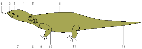
The internal structure of the lizardDigestive systemDigestive system
Mouth, oral cavity, pharynx, stomach, digestive glands, pancreas, liver, small and large intestines, cloaca - these are the sections of the digestive system of reptiles. In the mouth, saliva moistens the food, which facilitates its movement through the esophagus. Protein food is digested in the stomach under the action of gastric juice in an acidic environment. The ducts of the gallbladder, liver and pancreas open into the intestine. Here the digestion of food is completed and the absorption of nutrients into the blood occurs. Undigested food debris is expelled through the cloaca. excretory systemexcretory system
The excretory organs are the kidneys, ureters and bladder. SkeletonThe skeleton is completely bony. The spine is divided into five sections: cervical, thoracic, lumbar, sacral and caudal. The head is mobile due to the lengthening of the neck and the presence of two specialized cervical vertebrae. 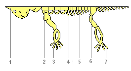
cervical consists of several vertebrae, with the first two providing head rotation in any direction. And this is extremely important for orientation with the help of the sense organs located on the head. Thoracic fixes the shoulder girdle through the chest and gives support to the forelimbs. Lumbar provides curves to the torso that help movement. Powerful sacral already consists of two vertebrae and the belt of the hind limbs becomes numb. long tail the department provides balancing movements of the tail. Since the oral cavity is no longer involved in gas exchange, the jaws have become elongated, more suitable for their main function - capturing food. Stronger jaw muscles attached to new projections on the skull made it possible to significantly expand the diet. Organ systemsRespiratoryRespiratory system
Breathing is only lung. The breathing mechanism of the suction type (breathing occurs by changing the volume of the chest), more advanced than that of amphibians. Conductive airways (larynx, trachea, bronchi) are developed. Internal walls and the septa of the lungs have a cellular structure. 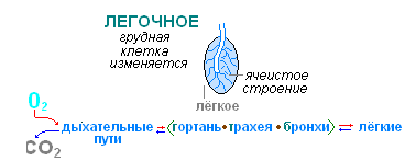 circulatoryCirculatory system
The heart is three-chambered, consists of two atria and one ventricle. An incomplete septum is developed in the ventricle. The large and small circles of blood circulation are not completely separated, but the venous and arterial flows are more strongly separated, so the body of reptiles is supplied with more oxygenated blood. The right atrium receives venous blood from all organs of the body, while the left atrium receives arterial blood from the lungs. When the ventricle contracts, its incomplete septum reaches the dorsal wall and separates the right and left halves. From the left half of the ventricle, arterial blood enters the vessels of the brain and anterior part of the body, from the right half of the venous blood goes to the pulmonary artery and then to the lungs. Mixed blood from both halves of the ventricle enters the trunk region. nervousNervous system
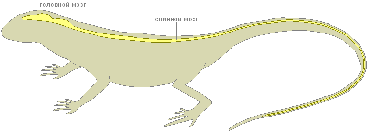
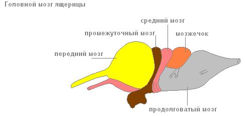
The brain is more developed, especially the forebrain hemispheres (responsible for complex instincts), the visual lobes and the cerebellum (coordinator of movements).  sense organsThe sense organs are more complex. The eyes of a reptile distinguish between both moving and stationary objects. The lens in the eyes can not only move, but also change its curvature. Lizards have movable eyelids. In the organs of smell, part of the nasopharyngeal passage is divided into olfactory and respiratory sections. 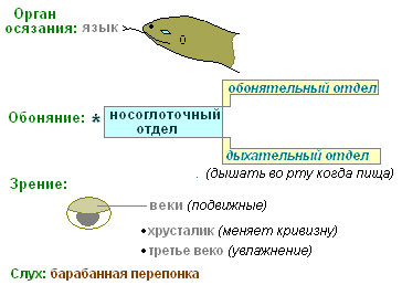 The internal nostrils open closer to the pharynx, so reptiles can breathe freely when they have food in their mouths. FertilizationLife appeared in water. Metabolic reactions take place in aqueous solutions. Water makes up the majority of any organism. Individual development The body needs a lot of water. Finally, without water, the movement of the spermatozoon and the fertilization of the egg are impossible. That is why, even in amphibians, fertilization and development are strongly associated with the aquatic environment. Overcoming this connection by reptiles is a big breakthrough in evolution. The transition to reproduction on land was possible only for animals capable of internal fertilization. Male reptiles have a special organ in the form of a permanent or temporary protrusion, with the help of which the seminal fluid from the testes is introduced into the female genital tract. This allows you to protect the sperm from drying out and provide them with the opportunity to move. Towards them, the eggs formed in the ovaries descend down the oviduct. In the same place, in the oviduct, the fusion of gametes occurs. 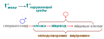 DevelopmentA fertilized egg is a large spherical yolk with a speck of the embryo on it. Going down the oviduct, the egg is surrounded by egg shells, of which the parchment shell is most pronounced in reptiles. It replaces the mucous membrane of amphibian eggs and protects the egg from external influences on land. In May - June, the female lays from 6-16 eggs in a shallow hole or mink. The eggs are covered with a soft fibrous leathery shell that prevents drying out. The eggs have a lot of yolk, the protein coat is poorly developed. Already at the beginning of the development of the embryo, an extra-embryonic bubble is formed from its tissues, which gradually surrounds the embryo from all sides. The embryo, along with the yolk, is suspended inside the egg. The outer shell of the bubble - serosa - creates antimicrobial protection. The inner shell - the amnion - limits the amniotic cavity, which is filled with fluid. It replaces the embryo with a water basin: it protects against concussions. 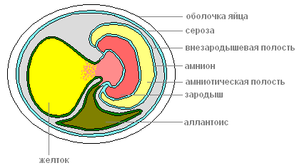 Cut off from the outside world, the fetus could suffocate and be poisoned by its own secretions. These problems are solved by another bubble - allantois, which is formed from the hindgut and grows into the first bubble. Allantois accepts and isolates all products of the excretion of the embryo, and returns the water back. In the walls of the allantois, blood vessels develop, which approach the surface of the egg and ensure the exchange of gases through the shells of the egg. Thus, allantois simultaneously plays the role of the germinal organ of excretion and respiration. All development takes 50-60 days, after which a young lizard hatches. The young cub is ready to live on land. It differs from an adult only in its smaller size and underdeveloped reproductive system. RegenerationVarious birds, small animals and snakes feed on lizards. If the pursuer manages to grab the lizard by the tail, then part of it is discarded, which saves it from death. 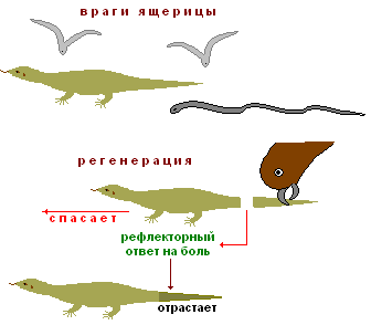 Tail drop is a reflex response to pain, it is carried out by breaking in the middle of one of the vertebrae. The muscles around the wound contract and there is no bleeding. Later, the tail grows back - regenerates. |
| Read: |
|---|

New
- List of sins with a description of their spiritual essence
- Prayer to the king of heaven comforter with accents
- Letter of Companion of the Prophet Muhammad
- The oldest monasteries in the world
- Ridiger, Mikhail Alexandrovich
- Request for Information
- What does many called but few chosen mean?
- Holy Apostles Peter and Paul - churches, icons, prayer
- Iconography of the Supreme Apostles Peter and Paul
- Kiev Caves Monastery (Lavra)


