Site sections
Editor's Choice:
- Reptiles structure. Sand lizard
- What gets men the most
- Traumatic brain injury - effects
- The main insect detachments table
- Military cap with his hands
- Household as a business
- How to fry the pollock in the pan
- Chaga mushroom: beneficial properties and contraindications
- Cooking a quick and tasty dinner: what can you cook at home inexpensively
- Muscle tension and muscle clamps for neurosis
Advertising
| Reptiles structure. Sand lizard |
|
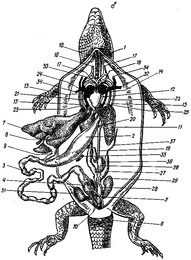
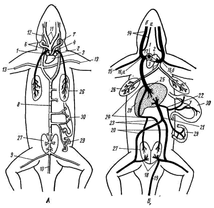
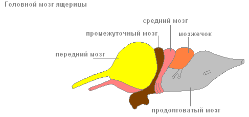
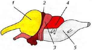
Repeat the general characteristics and classification of the Chord type. To study aromorphosis class Reptiles. Write in a notebook. Examine the structure of reptiles. Perform a summary in a notebook. Consider dry and wet preparations of different types of reptiles. To study the external and internal structure of reptiles using the example of a lizard (opening the lizard). In the album, make 5 drawings, indicated in printed manuals V (red tick). These printed manuals are stored in the laboratories department of biology and ecology, and with these manuals you work in practical classes. In the electronic training manual all the necessary pictures are placed at the end of the text. In a notebook, write down and learn the classification of modern and extinct reptiles. In the notebook draw and fill in table 1 .: Table 1. The variety of modern reptiles. Know the answers to test questions Topics: General characteristics of the type of Chord. Classification of Chord type. Features of the organization of reptiles. Systematic position, lifestyle, body structure, reproduction, value in nature and for humans Lizards. General characteristics of reptilesIn modern animal systematics Reptiles(Reptilia), or Reptiles, is a class in the Chordata type of the Vertebrate subtype (Vertebrata). Aromorphoses reptilesBasic Aromorphosis(Aromorphoses are major evolutionary changes leading to a general complication of the structure and organization of the body) The reptiles are as follows: 1. The emergence of germinal membranes, ensuring the development of the embryo in terrestrial conditions. Reptiles are amniotes, i.e. vertebrate animals whose embryos have embryonic membranes, ensuring the development of the embryo in the ground-air environment. 2. Progressive transformation of the skeleton and the formation of the chest. 3. The development of the brain, the appearance of the primordia of the cerebral cortex. 4. Differentiation of the muscular system (the appearance of intercostal muscles). 5. Differentiation of the respiratory tract and the appearance of cellular lung. 6. The development of incomplete interventricular septum of the heart. 7. The formation of the pelvic (secondary) kidney with the reverse absorption of substances. Reptiles- the first terrestrial vertebrates, which have lost their close connection with water. Among them are species that have returned to the water element for the second time. In the modern fauna there are about 6,600 species of reptiles. Reptiles appeared in the second half of the Carboniferous period of the Paleozoic era (about 300 million years ago). They are descended from more complex ancient amphibians, close to stegotsefalam. The highest flourishing and diversity of reptiles had in the Jurassic period of the Mesozoic era (195-135 million BP). The structure of reptilesThe structure of reptiles should be studied by example Lizards. Various species of lizards live throughout our country, except for the Far North. One of the most common and common species is the Sand lizard. Lacerta agilis (Chord type, Vertebrate subtype, Reptiles class, Scaly squad, Lizards suborder). This is a small animal, with a length of 15–20 cm with a tail. The lizard lives in dry, warmed places in the steppes, forests, mountains up to a height of 2.5 km. Brownish or greenish-brown color well hides it among the stones and grass. Lizards live in pairs, hiding at night in the burrows, under stones, under the bark of stumps. Here they are hiding from the fall to wintering. Body temperature is not constant. Lizards feed on insects and larvae of other invertebrates. In the warm season, several times during the summer, lizards lay in secluded places from 2 to 12 eggs. Care for the eggs do not show. Fully formed small lizards come out of eggs, and all their further development comes down to physical growth and puberty. Thus, the body of an elongated lizard consists of the head, neck, torso, tail and five-fingered limbs. Cover.The skin is dry, covered with horny scales, devoid of glands (protection against water loss). On the fingers are horn formations - claws. Growth is accompanied by molting (periodic change of the skin). Skeleton(Fig. 1) is completely ossified. It consists of an axial skeleton (spine), a skeleton of the head (skull) and a skeleton of limbs. Fig. 1. Skeleton of the Lizard 1 - the skull; 2 - clavicle; 3 - scapula; 4 - shoulder; 5 - brush; 6 - forearm bones; 7 - ribs; 8 - shin bones; 9 - foot; 10 - thigh; 11 - pelvic bones; 12 - the spine; 13 - sternum. General characteristics of reptiles The spine is divided into four divisions: 1. Cervical - includes eight vertebrae. The mobility of the head is provided by the first two cervical vertebrae - the atlas and the epistrophy, which form the joint. The first has the form of a ring and articulates with an unpaired condyle of the skull, the second is a dental process that enters the ring of the first vertebra and provides head rotation. 2. Thoracic-lumbar - consists of 22 vertebrae. It includes the thoracic region, where all vertebrae bear well developed ribs. The first five - long, attached to the sternum, forming the chest. The lumbar region is characterized by more developed transverse processes due to the growth of rudimentary ribs to them. 3. Sacral - includes two vertebra, to which the iliac bones of the pelvis are attached. 4. The tail - includes several dozen vertebrae, decreasing in size depending on the length of the tail. In case of danger, the lizard is saved from death by throwing off the tail (autotomy - a reflex act on pain), while one of the caudal vertebrae breaks in the middle. Later, the tail partially grows by regeneration. Skull almost completely ossified and consists of a large number of bones. The jaws are extended, which facilitates the capture and retention of prey. The large openings of the orbits are located on the top, not from the sides, like in fish. Connects to the spine with two condyles formed by the occipital bones. Limb skeletonincludes the belt and skeleton of free limbs, similar in structure to amphibians. Shoulder girdlerepresented by paired bones - shoulder blades, collarbone, crow bones (coracoids) and unpaired bone of the sternum. The shoulder belt is attached to the sternum. The skeleton of the forelimb consists of the upper arm (humerus), the forearm (radius and ulna), and the wrist (the wrist, metacarpus and phalanx of the fingers). Pelvic girdlerepresented by paired iliac, sciatic and pubic bones, fused together in the pelvis. It is attached to the two vertebrae of the sacral region. The skeleton of the hind limb consists of the femur (femur), tibia (tibia and tibia) and the foot (the bones of the tarsus, the metatarsus and the phalanges of the fingers). The bones of the extremities - ulnar and radial, as well as the large and small tibia do not grow together like in amphibians; foot and hand are shorter, fingers end in claws. Swimming membrane is absent. Muscular systemdeveloped and more differentiated in comparison with amphibians: there are rudiments of subcutaneous muscles, powerful chewing muscles (movement of the jaws), neck muscle groups (provide head movements), muscles of the extremities. Well-developed intercostal muscles that are involved in the mechanism of respiration. General characteristics of reptiles The internal structure of the lizard is shown in Figure 2. Nervous systemmore developed than amphibians. The brain of reptiles consists of five sections. Frontthe brain is represented by large hemispheres, divided into right and left lobes, of large size due to the accumulation of striped bodies. In the medulla, there is a primary arch (archipallium), which occupies most of the roof of the hemispheres, as well as the germ of a new cortex (neo-gallium). Progressive development of the forebrain ensures the emergence of complex behaviors. Intermediatethe brain is covered in front with the front, and behind - the middle brain. In the roof of the well developed parietal organ and epiphysis. Parietal organ its structure resembles the eye. It distinguishes a condensed transparent anterior part (similar to the lens) and a glassy back part consisting of sensory and pigment cells. It effectively perceives light stimulation and serves as a receptor for seasonal changes. Averagethe brain is enlarged due to the development of the visual centers. Cerebellumstrongly developed, the movements of reptiles are more intense and diverse. Oblongthe brain forms a bend in the vertical plane, also characteristic of all amniotes. Center of automatic motor activity and basic vegetative functions (respiration, digestion, blood supply). Eleven pairs of cranial nerves are moving away from the brain. Spinal cord enclosed in the spinal canal of the spine. Spinal nerves form the brachial and lumbar plexus. The sympathetic nervous system is well developed, represented by two nerve trunks located on the sides of the spine. Captions for Figure 2: 1 - esophagus, 2 - stomach, 3 - small intestine, 4 - large intestine, 5 - cloaca, 6 - vent of the cloaca, 7 - liver, 8 - gall bladder, 9 - pancreas, 10 - trachea, 11 - left lung , 12 - ventricle, 13 - right atrium, 14 - left atrium, 15 - right aortic arch, 15 - left aortic arch, 16 - right carotid artery, 17 - left carotid artery, 18 - left aortic arch, 19 - descending aorta , 20 - connecting the aortic arches to the dorsal aorta, 21 - the right pulmonary artery, 22 - the left pulmonary artery, 23 - the posterior vena cava, 24 - the venous sinus (weakly expressed in the lizard), 25 - the pulmonary ve on, 26 - left testicle, 27 - epididymis, 28 - seed tube, 29 - left kidney, 30 - infiltration of the seed tube into cloaca, 31 - bladder, 32 - right subclavian vein, 33 - spleen, 34 - left jugular vein, 35 - unpaired head vein, 36 - adrenal gland. General characteristics of reptiles
Fig. 2. The internal structure of the reptile (lizard, male). General characteristics of reptiles Sense organs.Reptilian senses (with the exception of snakes and some other species) are better developed than amphibians. eyes: they are more mobile. Their adaptation to vision at different distances (in many species at fairly large) is ensured not only by fish and amphibians, by moving the lens, but also, as in higher vertebrates, by changing its curvature. In addition to the eyelids, there is a blinking membrane. A number of species have parietal organassociated with the diencephalon and capable of perceiving light stimuli. Organ of hearing and balancerepresented by the middle and inner ear with three semicircular canals (organ of balance). The organ of hearing is adapted to the perception of sound stimuli in the air environment. The external auditory orifices are located on the head behind the eyes and tightened by a rounded eardrum that perceives sound vibrations. Oscillations of the membrane are transmitted to the auditory bone - stapes, - located in the middle ear cavity. The stirrup rests against the oval window leading to the cavity of the inner ear, transmitting to it vibrations of the eardrum. The lower part of the middle ear cavity is opened in the oropharynx using an auditory (Eustachian) tube to equalize pressure on both sides of the eardrum. Some progressive changes have occurred in the organ of hearing: the lower pouch slightly increased; the auditory ossicle better transmits the vibrations of the eardrum to the inner ear; in addition to the oval window, a second window appeared - round, which increases the mobility of the exolymph surrounding the labyrinth; eardrum is not located on the surface of the head, but in a small depression. AT touch language plays a big role. Organ of tasterepresented by pharyngeal taste bulbs. Organ sense of smell more differentiated and represented by olfactory bags, divided into olfactory (upper) and respiratory (lower) departments. An olfactory sink appears. There are olfactory passages with paired nostrils opening outwards. Respiratory system.The only respiratory organs in reptiles are the lungs, as their skin is impermeable to gases; The mucous membrane of the mouth, which plays a significant role in the breathing of amphibians, is used to perform this function by a few species of reptiles. For this reason, and especially due to the increased activity of reptiles compared to amphibians, the structure of the lungs and respiratory tract has become more complex. The lungs are organs of greater size and deeper located in the chest cavity than amphibians. The inner surface of them has increased significantly due to the development on it of many crossbars with a thicker network of blood vessels. The air enters the lungs after passing through the choanes, pharynx, larynx, and long trachea, the lumen of which is supported by cartilage rings. While passing through the trachea, the air is cleared of dust, its temperature approaches the body temperature. In amphibians, the trachea was in its infancy. The back end of the trachea is divided into two bronchi, in the walls of which there are also cartilaginous rings. Breathing in and out is more effective than in amphibians, due to the development of a reptile chest, which General characteristics of reptiles increases with inspiration and decreases with exhalation. The same mechanism is preserved in higher vertebrates - birds and mammals. Digestive system more differentiated than amphibians. The teeth are mainly focused on the jaws, their number is large, but they are of the same type and serve primarily to capture prey. Salivary glands are better developed than amphibians (in a number of species, these glands are poisonous). Reptiles begin the process of formation of a hard palate, which ends at the crocodiles. Due to the formation of a hard palate, the oral cavity is separated from the nasopharyngeal, which facilitates both breathing and food absorption. Throat, esophagus and stomach are well developed (especially in predators eating vertebrates). The liver (with gall bladder) and pancreas are more perfect than amphibians. The small intestine is significantly elongated; the colon is short and ends in a cloaca. Modern reptiles feed primarily on animals; the number of herbivorous species is insignificant. Circulatory systemclosed, two circles of blood circulation, three-chamber heart. In the ventricle of the heart there is an incomplete septum that prevents the complete mixing of arterial and venous blood. Three vessels depart from the ventricle: pulmonary trunk(from the right side of the ventricle) carries venous blood to the lungs. Right aortic arch(from the left side of the ventricle) carries arterial blood to the head (carotid arteries), fore limbs (subclavian arteries), then, merging with the left arch, forms the dorsal aorta. Left aortic arch(above the septum) carries mixed blood to all other organs of the body, merging into the dorsal aorta. Mixed (with high oxygen content) blood along the dorsal aorta is directed to the back of the body and to the hind limbs. The lizards maintain a sleepy duct that connects the carotid arteries with aortic arches. The metabolic rate remains low, so reptiles are cold-blooded animals. Small (pulmonary) circulationbegins with the pulmonary artery and ends with the pulmonary veins carrying arterial blood into the left atrium. Great Circle of Blood Circulationbegins with aortic arches and ends with the lower and two anterior hollow veins that carry blood to the right atrium. Excretory systemrepresented by paired oblong pelvic (metanephros) kidneys. From the kidneys depart ureters, opening into the cloaca. On the ventral side of the cloaca is the opening of the bladder. The mechanism of reabsorption in the renal tubules ensures the preservation of water in the body. The main end product of nitrogen metabolism is uric acid. General characteristics of reptiles The reproductive system.Separated. The sex glands are paired. Males have a copulatory organ. Fertilization is internal. Females lay large eggs with a high content of yolk. Outside the eggs are covered with a dense leathery shell. Reptiles are amniotes, i.e. vertebrate animals whose embryos have embryonic membranes, ensuring the development of the embryo in the ground-air environment. Developmentdirect. A young animal hatching from an egg that differs from an adult only in size, and all its development is reduced to physical growth and puberty. Reptiles are an important component of ecosystems, regulate the number of many invertebrates, serve as food for other animals. In a number of countries they are used by man for food, leather and shells are used for the manufacture of various products. The heart of the lizards is three-chambered, has two atria and one ventricle, divided into three parts: the venous cavity, the arterial cavity and the pulmonary cavity. Oxygen-poor blood enters the venous cavity from the right atrium, and oxygen-rich blood from the left atrium enters the arterial cavity. From the heart, blood escapes through the pulmonary artery, originating in the pulmonary cavity and two aortic arches extending from the venous cavity. All three lizards of the heart cavity communicate, but a muscle flap and biphasic ventricular contraction minimize blood mixing. Oxygen-poor blood flows from the venous cavity to the pulmonary, atrioventricular valve prevents it from mixing with oxygen-rich blood from the arterial cavity. Then the contraction of the ventricle pushes this blood from the pulmonary cavity into the pulmonary artery. The atrioventricular valve then closes, allowing oxygen-rich blood from the arterial cavity to enter the venous and leave the heart through the aortic arch. Thus, a three-chamber heart is functionally similar to a four-chamber heart. The paired left and right aortic arches behind the heart merge into the dorsal aorta. In reptiles there is a portal system of the kidneys - venous vessels of the tail and partially of the hind limbs lead directly to the kidneys. Thus, if injections of drugs excreted from the body by tubular filtration into the back half of the body are made, their concentration in the blood serum may be less than expected due to premature excretion in the urine. In the case of the introduction of nephrotoxic drugs, side effects may increase. However, there have been few studies of this effect, and their results indicate a rather insignificant role of the renal portal system in pharmacokinetics. Moreover, there are shunts in the system that carry blood from the portal system of the kidneys to the posterior vena cava, bypassing the kidney tissue. Lizards have a large abdominal vein, which lies along the inner surface of the middle of the abdominal wall on the bundle a few millimeters from the white line. When performing abdominal operations, this vein is avoided. However, if damaged, it can be ligated without complications. Digestive system of lizards Genitourinary system of lizards Nervous system of lizards Sense organs of lizards Respiratory system of lizards Musculoskeletal system of lizards Endocrine system of lizards CLOSED CLASS - REPTILIA THEME 12. OPENING OF THE LIZARD SYSTEMATIC POSITION OF THE OBJECT Subtype Vertebrates, Vertebrata MATERIAL AND EQUIPMENT For one or two students are needed: THE TASK Get acquainted with the features of the appearance of the lizard. To pay attention to the parts of the body, the structure of the integument, the external structure of the eyes, the external openings of the nostrils, the ear openings, etc. Perform an autopsy. Familiarize yourself with the general location of the internal organs; consistently consider the structure of individual organ systems, starting with the circulatory system. Make the following drawings: Additional task To examine, without sketching, under a microscope, a slice of lizard skin. APPEARANCE The body of the lizard is clearly divided into the head, neck, torso, tail and paired limbs - the front and rear (Fig. 71). Fig. 71. Appearance (A) and the area of the cloaca from below (B) of the Caucasian agama, male: The surface layers of the epidermis of the lizard skin (as well as of all other reptiles) harden: the cells gradually die off, filling with corneum - keratohyalin. Thickening of the stratum corneum occurs in small areas - scales, between which the stratum corneum is very thin (Fig. 72), so the flexibility of the skin (and the whole body) is maintained. The shape of the scales on different parts of the body of the same animal may vary significantly. Different species form, location and number of scales are usually more or less different, so these features are widely used in the systematics of reptiles.
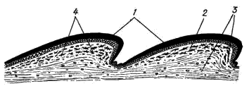
Fig. 72. Diagram of transverse incision of the skin of a lizard of the genus Lacerta.: The head of the agama is covered with small, irregularly shaped scales; some other lizards (for example, genera Lacerta, Eremias) have rather large horn shields on their heads, arranged in a strictly defined order. On the upper surface of the head, paired external nostrils are visible (Fig. 71, 1), opening into the oral cavity with so-called internal nostrils, or choanas (check by inserting a needle or bristle!). Eyes (fig. 71, 2) are covered with moving eyelids; in the back corner of the eye there is a blinking membrane. Behind the eyes are the ear openings (Fig. 71, 3), at some depth tightened by the eardrum. The elongated body of the agama is also covered with horny scales (Fig. 71, 5) —small, irregular-shaped scales on the dorsal side and rows of larger scutes on the belly. At the posterior end of the torso, on the border with the caudal region between the ventral plates, is a slit-like opening of the cloaca (fig. 71, 6). The tail scales of the Caucasian agama form double rings; other lizards have tail scales differently. The five-toed limbs of lizards, like other reptiles, end with horn formations - claws (Fig. 71, 4). The skin of lizards, like all reptiles, is dry, which is due to the absence of mucous glands. The skin glands are small in size and are located only on a few sites defined for this type. They secrete a thick, fat-like secret and carry special functions, most likely associated with leaving an odorous trail that facilitates the formation of pairs during reproduction. The agama clearly shows a group of such glands in the back of the abdomen; their secret in the form of "wax" plaque covers scales in this area. This accumulation of glands is especially well pronounced in males. OPENING 1. Place the lizard on its back in the wax bath and attach the limbs with pins to the bath. GENERAL TOPOGRAPHY OF INTERNAL BODIES Circulatory system. The heart (cor) is located on the ventral side of the anterior part of the chest cavity. Like amphibians, the heart of lizards is three-chambered: it consists of two atria - right and left (atrium dexter et atrium sinister; fig. 73, 1, 2) and one ventricle (ventriculus; fig. 73, 3).
Fig. 73. Diagram of the circulatory system of the Caucasian agama The ventricle of the heart is divided by an incomplete, so-called horizontal septum into two cavities: the smaller ventral (more precisely, ventrolateral), located down and to the right of the septum, and the larger dorsal (dorzolateral) - up and to the left of the septum. The left atrium opens into the left part of the dorsal cavity of the stomach, and the right atrium opens into the right part of the same cavity, in the region of the free edge of the septum. The dorsal cavity is divided into separate chambers by numerous muscle combs. One of them, the most developed, is the so-called vertical septum, which divides the dorsal cavity of the ventricle into two halves - left and right. Due to this structure in the ventricle of the reptile heart, there is no complete mixing of arterial and venous blood. With atrial contraction, arterial blood pushed out of the left atrium is collected mainly on the left side of the ventricular dorsal cavity; venous blood from the right atrium enters the right half of the dorsal part of the ventricle and, flowing around the edge of the horizontal septum, is collected in the ventral part of the ventricle. Only in the right half of the dorsal part of the ventricle, the arterial and venous blood is mixed. The arterial cone characteristic of amphibians is reduced in reptiles, and the main arterial trunks of the large and small circulation circulate independently of the ventricle. At the same time, unlike amphibians, in which three pairs of arterial trunks depart from the arterial cone, in reptiles only three unpaired vessels begin in the heart: the pulmonary artery and two (right and left) aortic arches. The pulmonary artery (arteria pulmonalis; fig. 73, 4) starts from the ventral (venous) part of the ventricle and soon divides into two branches that carry blood to the right and left lungs. On the pulmonary arteries moves venous blood. Oxygenated arterial blood through the pulmonary veins (vena pulmonalis; fig. 73, 5) returns to the heart. The right and left pulmonary veins merge into one unpaired vessel, which flows into the left atrium. The entire system of the vessels considered constitutes the small (pulmonary) circulation. Vessels of the systemic circulation also begin in the ventricle of the heart. The right aortic arch (arcus aortae dexter; Fig. 73, 6) departs from its left dorsal (arterial) part, and to its right, in the area of the free edge of the horizontal septum, the left aortic arch (arcus aortae sinister; Fig. 73, 7) . According to the place of discharge of these vessels in the ventricle, mainly arterial blood enters the right aortic arch, whereas mixed blood (arterial with venous impurity) enters the left artery. Both aortic arches bend around the heart and on the dorsal side behind it unite into an unpaired dorsal aorta (aorta dorsalis; fig. 73, 8), sending numerous vessels to various organs of the body. In the hindlimb, the dorsal aorta forks into two large iliac arteries (arteria iliaca; fig. 73, 9), which carry blood to the extremities, and the caudal artery (arteria caudalis; fig. 73, 10). The carotid arteries depart from the right aortic arch with a short, immediately split apart common trunk (arteria carotis; fig. 73, 11). Both carotid arteries, initially running parallel to the ascending branches of the aortic arches, carry blood to the head above the aortic arches turning up (down from the observer) and back each carotid artery sends a sleepy duct from itself (ductus caroticus; fig. 73, 12), which flows accordingly in the right or left aortic arch. All the vessels listed above are quite clearly visible on the freshly frozen lizard. If we carefully prepare the right aortic arch, then approximately in the middle between the place of its turn and the place of merging of the aortic arches, at the level of the posterior end of the heart, the subclavian arteries (arteria subclavia; fig. 73, 13) reaching the fore limbs can be seen. Thus, in reptiles, unlike amphibians, sleepy and subclavian arteries diverge asymmetrically - only from the right aortic arch. Due to this, blood richest in oxygen enters the head and forelimbs. ATthe enosal blood from the head is collected in large paired jugular veins (vena jugularis; fig. 73, 14), which, merging with the less visible subclavian veins from the forelimbs (vena subclavia; fig. 73, 15), form paired anterior hollow veins ( vena cava anterior dextra et vena cava anterior sinistra (Fig. 73, 16). The anterior vena cava falls into the venous sinus (sinus venosus; fig. 73, 17), which communicates with the right atrium. In lizards, the venous sinus, like most reptiles, is mild. From the back of the body, venous blood enters the heart in two ways. Veins carrying blood from the hind limbs form short paired portal veins of the kidneys (vena porta renalis; fig. 73, 18), with each of which merge branches of the divided unpaired tail vein (vena caudalis; fig. 73, 19). These vessels are usually able to be considered only on injected preparations. In the portal veins of the kidneys, blood enters the capillary system - the portal system of the kidneys. Most of the blood from the posterior part of the body goes through rather large paired pelvic veins (vena pelvica; fig. 73, 20; sometimes they are called the iliac veins - v. Iliaca), which merge to form an unpaired abdominal vein (vena abdominalis; fig. 73, 21) carrying venous blood to the liver. Venous blood from the intestine goes through several veins merging into an unpaired portal vein of the liver (vena porta hepatis; fig. 73, 22). In the liver or before entering it, the portal vein of the liver merges with the abdominal vein, and this general vessel immediately disintegrates into the system of hepatic capillaries. Consequently, as in amphibians, the portal system of the liver is formed by two veins: the abdominal and the portal liver. From the portal system of the kidneys, blood is collected into paired renal veins (vena renalis; fig. 73, 23), which merge into a large unpaired posterior vena cava (vena cava posterior; fig. 73, 24). The posterior vena cava penetrates the liver (not sending vessels into it) and flows into the venous sinus. From the portal system of the liver, blood is collected through the capillary system into the short hepatic vein (vena hepatica; fig. 73, 25), which flows into the posterior vena cava in the region of the anterior margin of the liver. Respiratory system. The respiratory tract of a lizard begins with the external nasal openings - nostrils. Next, the air through the nasal passage and internal nostrils - Hoan, enters the oral cavity. In the depth of the oral cavity, a few larynx (larynx) is located in front of the esophagus, consisting of three cartilage. It is equipped with special muscles and is connected with the hyoid apparatus. From the mouth, the inhaled air through the larynx enters the trachea (trachea; fig. 74, 4) - a rather long tube, in the walls of which there are ring-shaped cartilages that do not allow it to subside. The trachea runs along the neck and in the chest cavity, approximately at the level of the heart, divided into two short bronchus (bronchus) entering the lungs. The lungs (pulmones; fig. 74, 5) are thin-walled hollow bags. Compared to amphibian lungs in lizards, they have a more complex internal structure: their inner walls, in which capillaries branch, have a spongy structure, which markedly increases the total respiratory surface of the lungs. The lungs are the only respiratory organ of reptiles. The skin of these animals is dry, covered with horny scales and keratinized epithelium and is not involved in respiration. The act of breathing in lizards occurs by expansion and contraction of the chest under the action of special muscles.
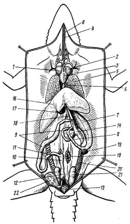
Fig. 74. The general arrangement of the internal organs of the Caucasian agama female: Digestive system. In the mouth there is a flat, tapering tongue anteriorly; It helps in seizing and swallowing prey. Many lizards and snakes tongue is thin and long, splitting at the end. It is very mobile, can protrude quite far from the mouth and also performs the function of the organ of touch: lizards and snakes feel the objects in front of them. In addition, when the tongue is retracted in the mouth, its tips fall into special grooves, equipped with sensory nerve endings - the Jacobson organ, which perceives chemical irritations from particles sticking to the tongue. In the posterior end of the oral cavity behind the laryngeal gap is the opening of the esophagus. The esophagus (oesophagus; fig. 74, 6) in the form of a muscular stretchable tube extends along the neck above the trachea and in the anterior part of the abdominal cavity flows into the stomach (gaster; fig. 74, 7). From the posterior end of the stomach, the duodenum (duodenum; fig. 74, 8) goes parallel to it and passes into the small intestine (ileum; fig. 74, 9). The border of the duodenal and small intestines is the first bowel bowel (the place where the bowel turns back). The small intestine makes several bends and passes into the large intestine (colon; fig. 74, 10). At the border of the small and large intestines there is a small blind growth - the anlage of the cecum (coecum; fig. 74, 11). The posterior part of the colon is the rectum (rectum; Fig. 74, 12). In lizards, the colon and rectum are separated by a poorly noticeable narrowing. The rectum opens into the cloaca (cloaca; fig. 74, 13) and through the cloacal fissure - out. An elongated compact pancreas is located between the stomach and the duodenum (pancreas; fig. 74, 14). Near the stomach, near the end of it, there is a small elongated reddish (on fresh material) spleen (lien; fig. 74, 15). The entire front part of the abdominal cavity (posterior to the heart) is occupied by a large liver with several lobes (hepar; fig. 74, 16). On its inner side is the gallbladder (vesica fellea; fig.74, 17). The bile duct which leaves it (ductus choledochus; fig. 74, 18) goes along the pancreas and flows into the beginning of the duodenum. The bile duct becomes more visible if you gently press the gallbladder with forceps and push a portion of the bile into the duct. Genitourinary system. In contrast to the previously studied classes, not the trunk (mesonephric), but the pelvic (metanephric) kidneys (ren; Fig. 75, 1; Fig. 76, 1) function in reptiles as adults. They are located in the back of the abdominal cavity and covered with bones of the pelvis. Along each kidney passes the ureter (ureter), opening into the cloaca. The ureters of lizards, like other reptiles, are formed simultaneously with the development of the metanephric kidney as thin-walled protrusions of the back of the wolf channels. The bladder departs from the abdominal wall of the cloaca in the form of a thin-walled blind outgrowth (vesica urinaria; Fig. 75, 2; Fig. 76, 2).
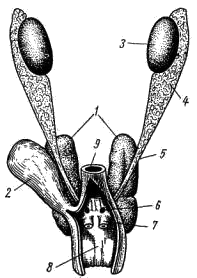
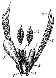
Fig. 75. The urinary system of the male Caucasian agama: The male sex glands - paired testes (testis; Fig. 75, 3) - are suspended from the mesentery in the dorsal back of the abdominal cavity. The testes with the help of the spermatozoa are closely connected with the epididymis (epididymis; fig. 75, 4), from which the seed tubes come (vas deferens; fig. 75, 5). Just before flowing into the cloaca, the seed ducts merge with the ureters and open in the cloaca with common openings (Fig. 75, 6). The appendages of the testis are remnants of the anterior part of the trunk (mesonephric) kidney, and the seed tubes are homologous to the excretory duct of this kidney, the wolf channel. Mullerian channels in males do not develop. In the side walls of the cloaca in the males there are two hollow outgrowths that can turn out through the opening of the cloaca to the outside. They play the role of aggregate organs. Fig. 76. Caucasian urogenital urinary system: The female sex glands are paired ovaries (ovarium; fig. 76, 4), suspended in the abdominal cavity on the mesentery and not directly connected to the excretory ducts. Ripened ovules fall out into the body cavity and then are captured by the funnel of the oviduct (Fig. 76, 6), which opens in front of the body cavity. Oviducts (oviductus; Fig. 76; 5), homologous to the Mullerian canals, open in the cloaca by separate (separate from the ureters) openings (Fig. 76, 7). The lower divisions of the oviducts in lizards are often extended and then get the name "uterus". Wolf channels in females are reduced. Reptiles are the first terrestrial vertebrates, part of the species has again passed to aquatic life. External structure(graphic image) Eggs of reptiles are large, rich in yolk and protein, covered with a dense parchment-like shell, developing on land or in the mother's oviducts. Water larva is absent. A young animal born out of an egg differs from an adult only in size. Dry skin is covered with horny scales and shields. 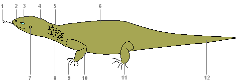
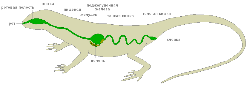
The internal structure of the lizardDigestive systemDigestive system
Mouth, mouth, pharynx, stomach, digestive glands, pancreas, liver, small and large intestines, cloaca - these are the parts of the digestive system of reptiles. In the mouth, saliva moistens the food, thus facilitating its movement along the esophagus. In the stomach under the action of gastric juice in an acidic environment, protein food is digested. In the intestines open ducts of the gallbladder, liver and pancreas. Here, the digestion of food is completed and the nutrients are absorbed into the blood. Undigested food residues are expelled through the cloaca. Excretory systemExcretory system
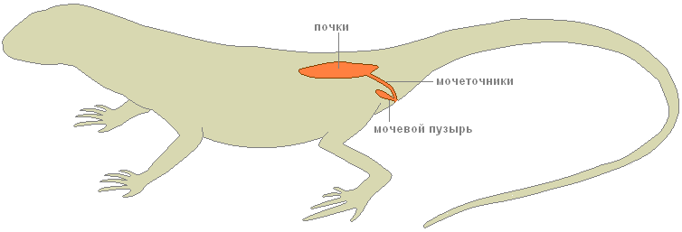
The organs of excretion are the kidneys, ureters and the bladder. SkeletonThe skeleton is completely bony. The spine is divided into five sections: cervical, thoracic, lumbar, sacral and caudal. The head is mobile due to the elongation of the neck and the presence of two specialized cervical vertebrae. 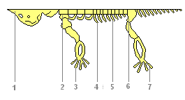
Cervical consists of several vertebrae, and the first two provide a turn of the head in any direction. And this is extremely important for orientation with the help of the sense organs on the head. Thoracic department through the rib cage fixes the shoulder girdle and gives support to the front limbs. Lumbar spine provides the bends of the torso helping movement. Powerful sacral section consists already of two vertebrae and the belt of hind limbs is numb on itself. Long tail the department provides tail balancing movements. Since the oral cavity is no longer involved in gas exchange, the jaws have become elongated, more suitable for its main function - food capture. Stronger jaw muscles, attached to new protrusions on the skull, made it possible to significantly expand the diet. Organ systemsRespiratoryRespiratory system
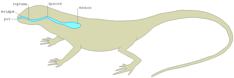
Breathing is only pulmonary. The breathing mechanism of the suction type (breathing occurs by changing the volume of the chest), more perfect than amphibians. Conducted respiratory tract (larynx, trachea, bronchi) are developed. The inner walls and partitions of the lungs have a cellular structure.  CirculatoryCirculatory system
The heart is three-chambered, consists of two atria and one ventricle. In the ventricle is an incomplete septum. Large and small circles of blood circulation are not completely separated, but the venous and arterial flows are demarcated more strongly, therefore the reptile body is supplied with more oxygenated blood. Venous blood from all organs of the body enters the right atrium, arterial blood from the lungs enters the left atrium. With the reduction of the ventricle, its incomplete septum reaches the dorsal wall and separates the right and left halves. From the left half of the ventricle, arterial blood enters the blood vessels of the brain and the anterior part of the body, from the right half venous blood flows into the pulmonary artery and further into the lungs. The trunk section receives mixed blood from both halves of the ventricle. NervousNervous system
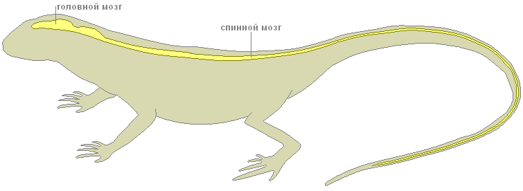
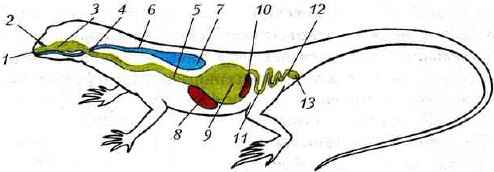
The brain is more developed, especially the forebrain hemispheres (responsible for complex instincts), the optic lobes and the cerebellum (coordinator of movements).  Sense organsThe sense organs are more complicated. Reptile's eyes distinguish between moving and non-moving objects. The lens in the eyes can not only move, but also change its curvature. In lizards, the eyelids are mobile. In the organs of smell, the part of the nasopharyngeal passage is divided into olfactory and respiratory parts. 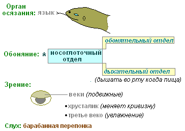 The internal nostrils open closer to the pharynx, so reptiles can breathe freely when they have food in their mouths. FertilizationLife appeared in the water. Metabolic reactions occur in aqueous solutions. Water makes up most of any organism. Individual development of the body requires a significant amount of water. Finally, without water, sperm movement and fertilization of the egg are impossible. That is why, even in amphibians, fertilization and development are strongly linked to the aquatic environment. Overcoming this connection with reptiles is a major breakthrough in evolution. The transition to breeding on land was possible only for animals capable of internal fertilization. The reptile males have a special organ in the form of a permanent or temporary protrusion, through which seminal fluid from the testes is introduced into the female genital tract. This helps to protect sperm from drying out and provide them with the ability to move. Towards them through the oviduct down the egg cells formed in the ovaries. There, in the oviduct, there is also a fusion of gametes. 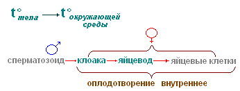 DevelopmentA fertilized egg is a large spherical yolk with a speck of germ on it. Going down through the oviduct, the egg cell is surrounded by egg shells, of which the parchment is most pronounced in reptiles. It replaces the mucous membrane of amphibian eggs and protects the egg from external influences on land. In May - June, the female lays from 6-16 eggs in a shallow hole or mink. Eggs are covered with a soft fibrous leathery shell that protects from drying out. The eggs have a lot of yolk, the protein shell is poorly developed. Already at the beginning of the development of the embryo, an extraembryonic bubble is formed from its tissues, which gradually surrounds the embryo from all sides. The embryo together with the yolk is suspended inside the egg. The outer shell of the bubble - serosa - creates antimicrobial protection. The inner shell - amnion - limits the amniotic cavity, which is filled with fluid. It replaces the embryo water pool: protects against shocks. 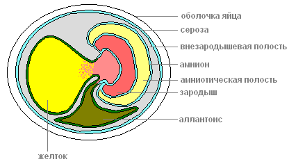 Cut off from the outside world, the embryo could suffocate and poison itself with its own secretions. One more bubble solves these problems - allantois, which is formed from the back intestine and grows into the first bubble. Allantois accepts and isolates all products of the embryo's release, and returns the water back. In the walls of the allantois, blood vessels develop, which approach the surface of the egg and provide for the exchange of gases through the shell of the egg. Thus, allantois simultaneously plays the role of the germinal organ of excretion and respiration. All development takes 50-60 days, after which a young lizard hatch. The young cub is ready to live on land. It differs from an adult only in its smaller size and underdeveloped reproductive system. RegenerationLizards feed on various birds, small animals and snakes. If the pursuer manages to grab the lizard by the tail, then part of it is discarded, which saves it from death. 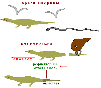 Tail drop is a reflex response to pain, it is performed by overlapping in the middle of one of the vertebrae. The muscles around the wound are reduced, and there is no bleeding. Later the tail grows back - regenerates. Features of the internal structure and life of reptiles are also considered on the example of a lizard. Nutrition and digestion. The digestive systems of reptiles and amphibians are similar in all major divisions (Fig. 143 and 144). These are the mouth, pharynx, stomach, intestines, cloaca. Fig. 143. The internal structure of the lizard (male): 1 - heart; 2 - trachea; 3 - lungs; 4 - gallbladder; 5 - the liver; 6 - the stomach; 7 - pancreas; 8 - small intestine; 9 - large intestine; 10 - the kidneys; 11 - bladder; 12 - cloacal opening; 13 - seed plants; 14 - seed lines In the mouth, saliva wets the food, which facilitates its movement along the esophagus. In the stomach under the action of gastric juice in an acidic environment, protein food is digested. In the intestines open ducts of the gallbladder, liver and pancreas. Here, the digestion of food is completed, the nutrients are absorbed into the blood.
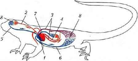
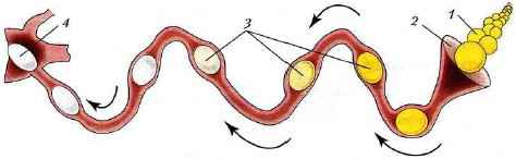
Fig. 144. Diagram of the digestive and respiratory systems of the lizard: 7 - mouth; 2 - nostrils; 3 - oral cavity; 4 - throat; 5 - esophagus; 6 - trachea; 7 - easy; 8 - the liver; 9 - the stomach; 10 - pancreas; 11 - small intestine; 12 - large intestine; 13 - cesspool Lizards eat mainly insects and worms, snakes - voles, mice, frogs. Some snakes on the front of the head have special sensitive pits - thermolocators capable of perceiving heat (infrared radiation) coming from a warm-blooded animal. Poisonous snakes kill prey from poisonous glands (located in the walls of the oral cavity), which runs down the poisonous teeth. Respiratory system. In connection with the appearance of the cervical region in the lizard, the airways are extended, through which air enters from the mouth into the lungs. The air is drawn in through the nostrils, enters the oral cavity, then into the larynx, then into the long tube - the trachea (see. Fig. 143). The trachea is divided into narrower tubes - bronchi, going to the lungs. Reptiles' lungs are more complex than amphibians. The walls of the lung cavity have many folds, where blood vessels branch out repeatedly. This increases the surface of their contact with air, enhancing gas exchange. Circulatory system. The heart is three-chamber, with an incomplete septum in the ventricle. Three large vessels emerge from it: the left and right aortic arches and the pulmonary artery (Fig. 145). Two arches of the aorta, beating around the heart, merge into one common vessel - the dorsal aorta.
Fig. 145. Diagram of the structure of the circulatory system of a lizard: 1 - heart; 2 - the carotid artery; 3 - left and right aortic arches; 4 - pulmonary artery; 5 - jugular (carries blood from the head) vein; 6 - intestinal vein; 7 - pulmonary vein; 8 - capillary network of internal organs Mixed blood flows through the body, like in amphibians, therefore reptiles have a non-constant body temperature, which depends on the ambient temperature. The pulmonary artery is divided into two branches that carry venous blood to the left and right lung. Here it is saturated with oxygen. Through the pulmonary veins, arterial blood enters the left atrium. In the ventricle, the blood is partially mixed, the richest in oxygen goes to the head, mixed - to all organs of the body, saturated with carbon dioxide - to the lungs. Nervous system. In reptiles, compared with amphibians, all parts of the brain are complicated and enlarged (Fig. 146). This is due to the more complex and diverse behavior of reptiles. Conditioned reflexes in them are formed faster than in fish and amphibians. The forebrain and cerebellum are especially enlarged, the medulla oblongata forms a bend, characteristic of all higher vertebrates. In addition to sight and smell, reptiles have a well-developed sense of touch.
Fig. 146. Diagram of the structure of the brain of a lizard: 1 - forebrain; 2 - diencephalon; 3 - the midbrain; 4 - the cerebellum; 5 - the medulla Excretory system. The excretory system of reptiles is the same as that of all terrestrial vertebrates. In the organs of excretion - the kidneys - the mechanism for the return of water to the body is enhanced. Therefore, the end product of metabolism in reptiles is not excreted in the form of liquid urine (as in amphibians), but in the form of uric acid in a pasty state in the cloaca, and then out. It does not require as much fluid for excretion of a mushy uric acid from the body as for excretion of liquid urine. Breeding bodies. In reptiles, like in other vertebrates, the reproductive organs of males are testicles, and in females - ovaries (Fig. 147). Fertilization in reptiles is internal. Seminal fluid enters the female genital tract when the cloaca of the male and the female come closer. The embryo in the fertilized egg develops even when the egg moves along the oviduct, covered with egg shells. They provide the embryo with water, protect against damage and shaking.
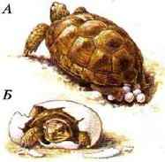
Fig. 147. Diagram of the structure of the oviduct of the lizard: 1 - ovary; 2 - oviduct funnel; 3 - promotion of a fertilized egg through the oviduct; 4 - egg covered in membranes in the foul place Reptiles lay eggs on the ground or in specially prepared cavities (Fig. 148). Some reptiles guard their clutch (for example, crocodiles); others, laying eggs, leave them (for example, turtles). Sometimes cubs develop in the mother's body. In these cases, egg production is occurring. For example, in the viper and in the viviparous lizard, the young hatch from the egg during its laying.
Fig. 148. Turtle laying eggs (A), and the release of a young turtle from an egg (B) Annual life cycle. Reptiles are widely distributed around the globe and are found in different climatic zones. However, being cold-blooded animals with a variable body temperature, they need heat from outside. Therefore, these animals are most numerous in tropical and subtropical zones of the globe. In the seasonal climate, where the warm summer is replaced by cold autumn and winter, the reptiles with the onset of adverse conditions go into shelters: burrows, caves, under tree roots, into the cellars of rural houses and forest huts. There the animals fall into a stupor - hibernation. In the spring, when the air and the surface of the soil are well warmed up, the reptiles come to the surface and switch to an active lifestyle. Reptiles are well adapted to living on land: they breathe in the lungs, they have internal fertilization, and the egg is covered with protective shells, providing the developing embryo with water and nutrients. Body temperature depends on the environment. At adverse times of the year, reptiles are held in shelters, falling into a stupor, in favorable periods are active. Exercises on the material
|

| Read: |
|---|
Popular:
What is the name of the golf course
|
New
- Name of IP for opening a farm
- Secrets of fishing: how to properly collect the bait
- General Class Amphibians
- How a man in love behaves
- Products that cause gas and bloating
- Why do sailors have a blue and white uniform?
- What does a hide-and-seek play teach a child?
- How to make a paper cap?
- How to pickle with pike caviar at home
- How to clean pike from scales and entrails


