Site sections
Editor's Choice:
- Muscles of the upper and lower extremities
- How to choose a spinning test
- Post-show work transplanting bees into clean, disinfected hives
- Recipes for barbecue with photos - cooking for men
- Economy Salad Recipes
- Pollock: cooking recipes in a pan
- The heaviest animal in the world
- How to draw a firefighter with pencil in stages
- NOD "Ecology" Subject: "In the mushroom kingdom, berry state
- How to make a paper out of paper
Advertising
| The structure of the muscles of the upper limb. Muscles of the upper and lower extremities |
|
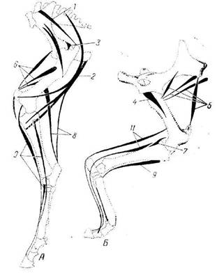
LECTURE №11. 1. Muscles of the shoulder girdle. 2. Muscles of the free upper limb. 3. The muscles of the pelvis. 4. Muscles of the free lower limb. OBJECTIVE: To know the topography and muscle functions of the shoulder girdle, shoulder, forearm, pelvis, hip and tibia. To be able to show these muscles on models, tablets and posters. 1. The muscles of the upper and lower extremities are divided into groups based on regional affiliation (topography) and the function they perform. The muscles of the upper limb can be divided into the muscles of the neck and the muscles of the free upper limb: shoulder, forearm and hand, the muscles of the lower limb - the muscles of the pelvic girdle (pelvis) and the free lower limb: thighs, shins and feet. The muscles of the shoulder girdle are located around the shoulder joint and provide it with a full range of movements (with the participation of certain muscles of the chest and back). All 6 muscles of this group begin on the bones of the shoulder girdle and attach to the humerus. 1) The deltoid muscle begins from the lateral third of the clavicle, acromion and spine of the scapula. Attached to the deltoid tuberosity of the humerus. The front part of the muscle bends the shoulder, the middle - removes, the back - extends the shoulder. 2) The supraspinatus muscle starts from the same fossa of the scapula, attaches to the large tubercle of the humerus. Shifts a shoulder, being a synergist of average bunches of a deltoid muscle. 3) The subosseous muscle starts from the same fossa of the scapula, attaches to the large tubercle of the humerus. Rotates the shoulder outwards. 4) The small round muscle starts from the lateral edge of the scapula and attaches to the large tubercle of the humerus. The synergist of the subostomy, i.e. rotates the shoulder outwards. 5) The big round muscle starts from the lateral edge and the lower corner of the scapula, and is attached to the crest of the small tubercle of the humerus. Pulls the shoulder down and back, while rotating it inward. 6) The subscapular muscle begins from the fossa of the same name and is attached to the small tubercle of the humerus and its crest. A synergist of the big round muscle and the broadest muscle of the back: the raised arm lowers, the lowered arm rotates inwards. 2. The muscles of the shoulder are divided into an anterior group — flexing muscles and posterior muscles — extensor muscles. The anterior group consists of 3 muscles. 1) The biceps muscle of the shoulder (biceps) has two heads: long and short. At the level of the middle of the shoulder, both heads are connected to the common abdomen. Bends the shoulder, forearm, rotating the latter outward (supination of the forearm). 2) The coraco-humeral muscle starts from the coracoid process of the scapula, is attached to the middle of the humerus from the medial side. Bends the shoulder and leads him to the body. 3) The shoulder muscle lies under the biceps muscle. Starts from the middle of the humerus, attached to the tuberosity of the ulna. Bends the forearm at the elbow. The posterior shoulder muscle group consists of 2 muscles. 1) The triceps muscle of the shoulder (triceps) occupies the entire posterior surface of the shoulder along its entire length. It has 3 heads. The lateral and medial heads begin on the humerus, and the long - on the subarticular tubercle of the scapula. Attached to the ulnar process. Extends the forearm, the long head extends the shoulder and leads him to the body (two-articular muscle). 2) The ulnus muscle is small. It starts from the lateral epicondyle of the humerus, attaches to the elbow bone and the back surface of the upper end of the ulna. Participates in the extension of the forearm. The muscles of the forearm are numerous and have a variety of functions. Most of them belong to the polyarticular, as it acts on several joints: elbow, radioulnar, wrist and distal joints of the hand and fingers. According to their position, they are divided into an anterior group — flexors and a posterior extensor. The front group consists of 7 flexors of the hand and fingers and 2 pronators, back - 9 extensors of the hand and fingers, and one muscle instep. The front muscles of the forearm form 2 layers: superficial and deep. The surface layer includes 6 muscles. 1) The humeral muscle starts from the humerus above the lateral epicondyle and attaches to the distal end of the radius. Bends the forearm, sets it and the hand in the middle position between supination and pronation. 2) A round pronator begins, like all remaining superficial muscles, from the medial numfix of the humerus. Attached to the middle third of the radius. Penetrates and flexes the forearm at the elbow. 3) The radial flexor of the wrist is attached to the base II of the metacarpal bone. Bends and partially permeates the brush. 4) The long palmar muscle is attached to the palmar aponeurosis. It strains the palmar aponeurosis, participates in the flexion of the hand. 5) The surface flexor of the fingers is wide, covered in front by the muscles described. It is divided into 4 long tendons, which are attached each with two legs to the base of the middle phalanges of II-V fingers. Bends the middle phalanges of these fingers and hand. 6) The elbow flexor of the wrist is attached to the pea-shaped bone. Bends the brush and participates in its cast. The deep layer of the forearm forearm includes 3 muscles. 1) The long flexor of the thumb begins from the radius, attached to the distal phalanx of the thumb. Bends the distal phalanx of the thumb, participates in the flexion of the hand. 2) The deep flexor of the fingers starts from the ulna and attaches to the bases of the distal phalanges of II-V fingers. It bends the distal phalanxes of II-V fingers and the whole hand. 3) Square pronator is located in the area of the distal ends forearm bones. It starts from the medial edge of the body of the ulna, attached to the lateral edge and the front surface of the radial bone. The main pronator of the forearm (rotates the forearm inward). The muscles of the back group of the forearm unbend the hand and fingers, rotate the forearm outwards (supin), together with the muscles of the shoulder participate in the extension of the forearm. They also form 2 layers - superficial and deep. Superficial muscles begin from the lateral epicondyle of the humerus, deep - from the bones of the forearm, mainly from the ulna. The surface layer of the back group of the forearm includes 5 muscles. 1) The long and short radial extensors of the wrist are attached: long - to the second metacarpal bone, short - to the third metacarpal bone. Extend the brush. 2) The extensor of the fingers is attached by four tendons to the phalanges of II-V fingers. Extends fingers and hand. 3) The elbow wrist extensor is attached to the base of the V metacarpal bone. Extends and leads the brush. 4) The extensor of the little finger is attached to the phalanxes of the V finger. Extends the little finger. The deep layer of the back group of the forearm also includes 5 muscles. 1) The forearm support is attached to the radius. Rotates the forearm outward. 2) The long muscle that retracts the thumb of the hand is attached to the base of the metacarpal bone. Removes the thumb and the whole brush. 3) The short and long extensors of the thumb are attached respectively to the base of the I and II phalanges of the thumb. Extend the thumb of the brush, removing it. 4) The extensor of the index finger is attached to the proximal phalanx of the index finger. Extends the index finger (“pointing” mouse). The muscles of the hand are located mainly on the palmar side. They are divided into 3 groups: lateral, middle and medial. Lateral group - muscles of an eminence of a thumb (tenar) includes 4 short muscles: 1) short thumb flexor; 2) short muscle, retracting the thumb; 3) the muscle leading the thumb; 4) muscle opposing the thumb. Medial group - muscles of an eminence of a little finger (hypothenar) also includes 4 short muscles: 1) short palmar muscle; 2) the muscle that removes the little finger; 3) short little flexor; 4) muscle opposing little finger. Medium muscle group includes: 1) worm-like muscles (there are four of them), flex the main phalanges and unbend the middle and distal phalanges of the II-V fingers; 2) interosseous muscles: palmar (there are 3) - they bring II, IV and V fingers to the middle (III) and back (4) - I, II, IV fingers are removed from the middle finger. 3. The pelvic muscles, starting on the bones of the pelvis of the Iv-pillar, surround the hip joint and attach to the upper end of the femur. The muscles of the pelvis are divided into internal and external groups. The internal (front) group of muscles of the pelvis includes 4 muscles. 1) The iliopsoas muscle consists of two muscles that are connected into one: the large lumbar and ileal. The first begins from the XII thoracic and all lumbar vertebrae, the second from the iliac fossa. Coming behind the inguinal ligament through the muscle lacuna in the thigh area, the iliopsoas muscle attaches to the small skew of the femur. Bends the thigh and turns it outwards, with a fixed thigh tilts the pelvis, along with the body forward. 2) The small lumbar muscle is unstable (absent in 40% of cases). It starts from the sides of the bodies of the XII thoracic and I lumbar vertebrae, attached to the iliac fascia and the crest of the pubic bone. Tightens the iliac fascia. 3) The pear-shaped muscle begins from the pelvic surface of the sacrum, emerges from the pelvic cavity through the large sciatic opening and attaches to the tip of the greater trochanter of the femur. Rotates the thigh outward. 4) The internal obturator muscle starts from the inner surface of the pelvic bone, the obturator membrane, emerges from the pelvic cavity through the small sciatic hole, attaches to the trochanteric fossa of the femur. Rotates the thigh outwards. The external (posterior) group of muscles of the pelvis includes 8 muscles. 1) The gluteus maximus muscle reaches its greatest development in man due to erect position. It starts from the outer surface of the wing of the Ilium, the sacrum and the coccyx, and is attached to the gluteal tuberosity of the femur (third skewer). Extends the thigh, rotates it outward, and when standing, fixes the pelvis and torso, also serves as a pillow. 2) The gluteus maximus muscle is under the previous one. It starts from the gluteal surface of the ilium, attaches to the greater trochanter of the femur. Pulls the hip, front beams rotate the thigh inward, rear - out. 3) The gluteus maximus muscle lies deeper than the gluteus maximus muscle. It has a similar beginning, attachment and function. 4) The external obturator muscle starts from the obturator membrane and the external circumference of the obturator opening, attached to the trochanteric fossa of the femur. Rotates the thigh outwards. 5) The square muscle of the hip begins from the sciatic knob, attached to the greater trochanter and intertrochanter crest of the femur. Rotates the thigh outwards. 6) The upper and lower twin muscles are located above and below the tendon of the internal obturator muscle. Begin: the first from the ischial spine, the second from the ischial tuber. Attached to the trochanteric fossa of the femur. Rotate the thigh outwards. 7) The retractor of the wide fascia begins from the superior anterior iliac spine, continues downward, intertwines into the ileal-tibial tract of the wide fascia of the thigh and stretches (strains) this fascia, participating in the flexion of the thigh. 4. The thigh muscles perform static and dynamic functions when standing, walking. Like the muscles of the pelvis, they reach maximum development in humans due to erect position. The muscles of the thigh are divided into 3 groups: anterior (hip flexors), hind (extensors of the thigh) and medial (leading thigh). The anterior group includes two muscles. 1) The tailor muscle is one of the longest muscles in the human body (about 60 cm). It starts from the upper anterior iliac spine and attaches to the tibial tuberosity, fascia of the tibia. Bends the thigh and shin, rotates the hip outwards, and shin - inside. 2) The quadriceps muscle of the thigh (quadriceps) is the most voluminous and strong muscle in the whole body (weighing up to 2 kg). It consists of four heads: the rectus femoris muscle, the lateral, medial and intermediate wide muscles of the femur. Connecting together, the heads of the common tendon are attached to the base and the side edges of the patella. Bends the lower leg, rectus muscle bends the thigh. The posterior thigh muscle group includes 3 muscles. 1) The biceps muscle of the thigh begins with a long head from the sciatic mound, a short head from a rough line. Together, both heads are attached to the head of the fibula. Extends the thigh, flexes the shin, rotates the bent shin out. 2) The semi-tumble muscle starts from the sciatic tuberosity, attaches to the tibial tuberosity. Bends the thigh, bends the shin, rotates the bent shin inward. 3) The semi-membranous muscle starts from the sciatic tubercle and is attached by a flat tendon of three bundles to the posterolateral surface of the medial condyle of the tibial bone. The medial thigh muscle group includes 5 muscles that are not joined together. only position, but also a common function: they lead the thigh: 1) the comb muscle; 2) the thin muscle (the muscle of the "virginity"); 3) the long adductor muscle; 4) the short adductor muscle; 5) the large adductor muscle. All these muscles start from the pubic and partially from the sciatic bone, attached (with the exception of the thin muscle) to the rough line of the femur. The thin muscle is attached to the tibial tuberosity and is involved not only in bringing the thigh, but also in flexing the tibia and turning it inwards. The calf muscles surround both tibial bones, forming the anterior, posterior, and lateral groups. The bones of the lower leg and the interosseous membrane delimit the anterior and posterior muscle groups. The front group - extensors of the foot - includes 3 muscles. 1) The anterior tibial muscle extends the foot in the ankle joint, lifts its medial edge (supination). 2) The long extensor of the fingers begins from the proximal ends of the bones of the tibia and is attached to the phalanges of II-V fingers. A small bundle is separated from the lower part of the muscle - the third peroneal muscle, which is attached to the V metatarsal bone. Extends fingers and foot, raises the lateral edge of the foot. 3) A long extensor of the big toe extends the thumb and foot. The back group - flexors of the foot - includes 6 muscles. 1) The triceps muscle of the lower leg is formed by three heads, of which two (superficial) constitute the gastrocnemius muscle, and one (deep) muscle is the soleus. Both muscles end with a heel (Achilles) tendon attached to the heel puff. Bends the shin, bends and rotates the foot outward. 2) Plantar muscle non-permanent. Tightens the capsule of the knee joint, is involved in the flexion of the leg and foot. 3) The popliteal muscle lies in the bottom of the popliteal fossa. Bends the calf, turning it inwards. 4) The posterior tibial muscle is located deep on the back the surface of the lower leg, between the long flexor of the fingers (medially) and long flexor of the big toe (lateral). Flex foot, brings her and supiniruet (rotates out). 5) A long flexor of the fingers is attached to the distal phalanges of II-V fingers. Bends these phalanges, foot, turning it out. 6) The long flexor of the big toe is attached to the distal phalanx of the thumb. It bends the big toe, participates in the bending, supination and adduction of the foot, strengthens the longitudinal arch of the foot. Lateral muscle group of the lower leg, raising the lateral margin foot, includes 2 muscles. 1) Long fibular muscle. 2) Short fibular muscle. Both of these muscles start from the fibula, their tendons pass to the foot behind the lateral ankle and attach the first to the base of the I-II metatarsal bones, the medial sphenoid bone, the second to the V metatarsal bone. Bend the foot, produce pronation, strengthen the transverse and longitudinal arches of the foot. The muscles of the foot are divided into the muscles of the back and plantar surfaces. The muscles of the back of the foot include two short muscles involved in the extension of the toes of the foot: 1) a short extensor of the fingers; 2) a short extensor of the big toe. On the sole of the foot, as in the hand, there are 3 muscle groups. The medial group (muscles of the big toe) includes 3 muscles: 1) a muscle that removes the big toe; 2) a short flexor of the big toe; 3) a muscle that causes the big toe. The lateral group (muscles of the little finger of the foot) also includes 3 muscles: 1) the muscle that removes the little finger of the foot; short flexor of the little toe of the foot; 3) muscle opposing little finger (non-permanent). The middle group of muscles of the sole of the foot includes the following muscles: 1) a short finger flexor; 2) a square muscle of the sole (accessory flexor); 3) four worm-like muscles; 4) interosseous muscles: the plantar - three, and the back muscles - four. The deepest of the short muscles of the foot are located in the gaps between the metatarsal bones of the foot. The muscles of the upper limb are divided into the muscles of the shoulder girdle and the muscles of the free upper limb: shoulder, forearm and hand. Shoulder muscles The back muscle group of the forearm. 2. Medial group - the muscles of the hypotenar: short palmar muscle; the muscle that removes the little finger; short flexor of the little finger; muscle opposing little finger. These muscles fix the little finger and carry out its flexion, abduction and opposition .______________________________________________________________ Another variant!!! The structure includes: Leather. Muscles Bone skeleton. Blood vessels. Bundles Muscle Anatomy Fibers are divided into two types. The first is the musculature shoulder girdleto the second - free part. Classification is carried out depending on the tasks and location. Muscles of the upper limbs in areas of the shoulder girdle are divided into deltoid, supra-subarate, small and large round, as well as subscapularis fibers. The composition of the shoulder girdle includes muscles of the hand, shoulder and forearm. Large round fibers They have an oblong flat shape. Start from the back of the lower corner of the shoulder blade. These muscles of the upper limbs are fixed on a small tubercle in the humerus (on the crest). The back calving adjoins the broad fibers of the back. The large, round muscles of the upper limbs, while contraction, pull the shoulder backwards, turning it inwards. As a result, the arm returns to the body. Deltoid fiber They are presented in the form of a triangle. Under the lower part of this muscle of the upper extremities are the subdeltoid bags. The fibers cover the shoulder joint completely and the shoulder musculature locally. The deltoid muscle includes large bundles converging on top. They are divided according to the tasks. The rear pull back the hand, the front - forward. The fibers begin from the axis of the scapula (lateral end) and part of the clavicle. Fixation area - deltoid tuberosity in the humerus. The deltoid muscles of the upper limbs move the shoulders outwards until they assume a horizontal position. Small round fibers They make up the elongated circular muscle. The front part of it is covered with deltoid fibers, the back part - with large round ones. The muscle begins from the scapula, slightly below the subshell fibers, to which its upper surface abuts. Attach the segment to the site on the humerus of the humerus and the capsule of the joint (to the back of it). The muscle turns the shoulder outwards, retracts and retards the joint capsule. Supraspinatus fibers They form a triangular muscle. It is located in the supraspinous fossa under the trapezoidal segment. The place of fixation is the back of the capsule of the shoulder joint and the platform on the large tubercle of the bone. The muscle begins on the surface of the fossa. With the reduction of the fibers, the shoulder rises and the joint capsule is pulled away, which prevents pinching. Undercut fibers They formed a triangular wide flat muscle. The fibers are located in the subscapular fossa. At the site of attachment there is a tendon bag. The muscle begins on the subscapular fossa, and ends in the small tubercle in the humerus and on the front of the joint capsule. By reducing the fibers, the shoulder rotates inward. Subostine fibers They form a flat triangular muscle. The segment is located in the posterior fossa. The beginning of the fibers is located on its wall and rear scapular part. It is fixed to the capsule in the shoulder joint and to the middle platform on a large bone tuberosity, under which the tendin bag is located. Contraction, the muscle turns the shoulder outwards, allows you to withdraw a raised arm, pulls the joint capsule. Shoulder muscles It is divided into two groups. The front bends, and the back bends the shoulder and forearm. The first group includes biceps, brachial and coracoid muscles. The composition of the second section consists of the triceps and elbow muscles of the human upper limbs. Double headed fibers They form a spindle-shaped round muscle. In its composition there are two heads: a short one, which performs the cast of a hand, and a long one, which produces a lead. The latter starts from the supra-articular tubercle of the scapula. The short head departs from the coracoid process. In the place of their connection the belly is formed. It attaches to the tubercle on the radius. In the medial direction are several fibrous bundles. They form a laminar process - aponeurosis. Then it goes into the humeral fascia. The objectives of the biceps muscle are rotating outward and flexing the forearm at the elbow. Beak fiber They form a flat muscle. It is covered with a short head of the two-headed segment. The klyuvovidnye muscles of the upper limbs of a person begin at the top of the same process of the scapula. Attach the fibers below the center of the medial part of the humerus. Due to their reduction, the shoulder rises, the hands are brought to the midline. Shoulder fibers They formed a wide spindle-shaped muscle. The front and outer surfaces of the shoulder bone are its beginning. The fixation is made to its tubercle and the capsule of the elbow joint. The fibers are completely located in the lower shoulder (on the front side) under the biceps muscle. Elbow segment This muscle has a pyramidal shape. Its beginning is the lateral epicondyle of the shoulder bone. Fibers are attached to the back of the body of the ulna and the process of the same name. When shrinking, the muscle extends the forearm. She also coordinates the capsule pulling off at the elbow. Triceps fiber They form a long muscle. It consists of 3 heads: medial, lateral and long. The beginning of the latter is the subarticular scapular tubercle. The lateral head departs from the posterior-lateral part of the shoulder bone, the medial head - from the posterior surface. Elements are connected in a spindle-shaped abdomen. It subsequently turns into a tendon. The abdomen is fastened to the joint capsule and the process of the elbow. With the reduction of fibers, the forearm is unbent, the arm is pulled back and the shoulder is brought to the body. The muscle is located from the olecranon to the scapula. Forearm fibers They form two muscle groups: anterior and posterior. In each of them there are fibers of the deep and surface layer. The latter in the anterior group include the flexors of the hand (ulnar and radial) and the fingers, the shoulder-beam segment, the circular pronator. The department also includes long palmar muscles. In the deep layer there are square pronator, flexors: long thumb and deep finger. The superficial muscles of the posterior group include the elbow, short, and long radial extensors of the wrists, the finger, and the little finger. In the deep layer of the department there are an instep, muscles, abductor and extensors of the thumb (short and long), an extensor for the index finger. Musculature of the hand Muscles are located on the palmar surface. Fibers are divided into several groups: medium, medial, lateral. On the back of the surface of the hand are the intercostal muscles of the same name. In the lateral group there are fibers that correct the movement of the thumb: opposing, leading, flexors and discharging. The medial part includes the short palmar muscle and little finger musculature. The latter includes a short flexor, leading and discharging fibers. In the middle group there are worm-shaped, palmar and dorsal interosseous elements. The muscles acting on the joints of the limbs are perpendicular to the axis of movement in the joints. The greatest number of muscles acts on the multiaxis joints - shoulder and hip. The flexors lie inside the corners of the joints, and their abdomens are higher than the joints that they flex. The extensors also come up on top and lie on top of the angle of the joint. Adductors on the thoracic limbs are abdomens on the inner surface of the scapula and act through the shoulder joint. Adductors on the pelvic limb lie abdomens on the femur, but act through the hip joint. Abductors are abdomens on the outer side of the joint and above the joint through which they act. Rotators go obliquely to the axis of the joint. Muscles, as well as bones, are more developed on the pelvic limbs than on the pectoral limbs, since when the animal moves, the main work is done by the pelvic limbs. The muscles of the chest limbs (Fig. 22, 23, 24). The shoulder joint is affected by the extensors of the shoulder joint — the forearm, the biceps of the shoulder, and the brachiocephalic muscle. The flexors of the shoulder joint are large and small round muscles, the deltoid muscle, the latissimus dorsi muscle, and the long head of the triceps muscle. The limb abductor is an abnormal muscle. Adductors of the limbs - subscapularis and coraco-brachial muscles. Adductors help the pectoral muscles (from the group of muscles of the shoulder girdle). The deltoid and small round muscles, in addition, rotate the limb outward (supination), the large round and latissimus dorsi muscle rotate the limb inward (pronation). On the elbow joint, as in all subsequent ones, there are only two groups of muscles - the extensors and the flexors. The extensors of the elbow joint include the triceps muscle of the shoulder (very powerful), the elbow muscle, and the fascia tensioner of the forearm. They are helped by flexors of the wrist and fingers. The flexors of the elbow joint include: the biceps muscle of the shoulder and the inner shoulder muscle. They are helped by the radial extensor of the wrist and the general finger extensor. The extensors of the wrist are: the radial extensor of the wrist and the long abductor of the thumb; they are helped by finger extensors. Fig. 24. Layout of the extensors (A) and flexors (B) of the chest limbs: 1 - extensors of the shoulder joint; 2 - abductor of the shoulder joint; 3 - flexors of the shoulder joint; 4 - shoulder joint adductor; 5 - extensors of the elbow joint; b —- flexors of the elbow joint; 7 - wrist extensors; 8 - wrist flexors; 9 - finger extensors; 10 - finger flexors. Wrist flexors include: radial and ulnar flexors of the wrist and ulnar extensor of the wrist (in ungulates); they are helped by finger flexors. The fingers are affected by common and lateral finger extensors and superficial and deep finger flexors. In addition, the intercostal muscles, which in hoofed animals (especially in horses) have become powerful tendons, affect the knot joint. The muscles of the pelvic limb (Fig. 22, 25). Movement in the hip joint is carried out by the extensors, flexors, adductors, abductors and rotators. The extensors include the powerful gluteus muscles and the so-called hip femur group, which includes the biceps of the thigh, the semimembranosus, semitendinosus, and the square muscles of the thigh. By the flexor belongs lumbar-iliac muscle; she is helped by the tensor of the wide fascia of the thigh, the tailor's, the comb-like muscles, and the straight head of the quadriceps of the thigh. The pelvic limb adductors are slender and adductor muscles, the abductors are the gluteus maximus muscle. Rotators include locking and twin muscles.
Fig.25. The layout of the extensors (A) flexors (B) pelvic limb: 1 - gluteus muscles; 2 - biceps muscle of the thigh; 3 - hip joint abductor; 4 - hip joint adductor; 5 - hip flexors; b - extensors of the knee joint; 7 - knee flexors; 8 - extensors of the tarsal, or hock, joint; 9 - flexors of the tarsus, or hock, joint; 10 - finger extensors; 11 - finger flexors. The extensor of the knee joint is the quadriceps muscle of the thigh, and the biceps of the thigh helps it. The knee flexors include: biceps of the thigh, semitendinosus and gastrocnemius muscles; they are helped by the popliteal muscles, the main function of which is rotational. At the tarsus joint there are extensors in the composition: the triceps of the calf (mainly the gastrocnemius muscle) and the biceps of the thigh; it helps the surface flexor of the fingers. The anterior tibial muscle flexes the hock; she is assisted by a long finger extensor and the third peroneal muscle. The fingers are affected by long, lateral and short finger extensors and superficial and deep finger flexors. As with the pectoral limb, there is an interosseous muscle acting on the gang joint. |
| Read: |
|---|
New
- Reptiles structure. Sand lizard
- What gets men the most
- Traumatic brain injury - effects
- The main insect detachments table
- Military cap with his hands
- Household as a business
- How to fry the pollock in the pan
- Chaga mushroom: beneficial properties and contraindications
- Cooking a quick and tasty dinner: what can you cook at home inexpensively
- Muscle tension and muscle clamps for neurosis


